Urolithiasis, commonly known as kidney stones, causes blockage in the urinary tract, often leading to hydronephrosis, a condition characterized by the swelling of a kidney due to urine buildup. Early diagnosis and treatment are crucial to prevent permanent kidney damage and alleviate pain. Explore the rest of the article to learn how you can recognize symptoms and effective treatment options.
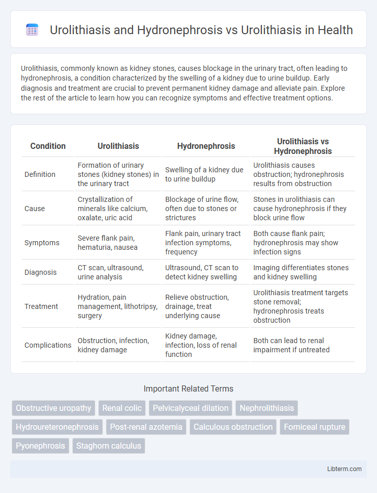
Table of Comparison
| Condition | Urolithiasis | Hydronephrosis | Urolithiasis vs Hydronephrosis |
|---|---|---|---|
| Definition | Formation of urinary stones (kidney stones) in the urinary tract | Swelling of a kidney due to urine buildup | Urolithiasis causes obstruction; hydronephrosis results from obstruction |
| Cause | Crystallization of minerals like calcium, oxalate, uric acid | Blockage of urine flow, often due to stones or strictures | Stones in urolithiasis can cause hydronephrosis if they block urine flow |
| Symptoms | Severe flank pain, hematuria, nausea | Flank pain, urinary tract infection symptoms, frequency | Both cause flank pain; hydronephrosis may show infection signs |
| Diagnosis | CT scan, ultrasound, urine analysis | Ultrasound, CT scan to detect kidney swelling | Imaging differentiates stones and kidney swelling |
| Treatment | Hydration, pain management, lithotripsy, surgery | Relieve obstruction, drainage, treat underlying cause | Urolithiasis treatment targets stone removal; hydronephrosis treats obstruction |
| Complications | Obstruction, infection, kidney damage | Kidney damage, infection, loss of renal function | Both can lead to renal impairment if untreated |
Overview of Urolithiasis
Urolithiasis refers to the formation of stones in the urinary tract, primarily composed of calcium oxalate, uric acid, or struvite, causing obstruction and pain. This condition can lead to complications such as hydronephrosis, where urine accumulates in the kidney due to blockage, causing renal swelling and possible damage. Early diagnosis using imaging techniques like ultrasound and CT scans is crucial for effective management and prevention of kidney function impairment.
Understanding Hydronephrosis
Hydronephrosis is the swelling of a kidney due to a build-up of urine caused by an obstruction such as urolithiasis, which refers to the formation of kidney stones. In urolithiasis-related hydronephrosis, kidney stones block urine flow, leading to increased pressure and kidney tissue damage if untreated. Early diagnosis and treatment are critical to prevent complications like chronic kidney disease and preserve renal function.
Pathophysiology of Urolithiasis
Urolithiasis involves the formation of calculi within the renal system due to supersaturation of urine with stone-forming solutes such as calcium, oxalate, uric acid, and cystine, leading to crystal nucleation, growth, and aggregation. Hydronephrosis occurs when these obstructive calculi impair urinary flow, causing dilation of the renal pelvis and calyces, increased intrarenal pressure, and potential renal parenchymal damage. Understanding the pathophysiology of urolithiasis highlights the critical role of urinary pH, inhibitor deficiencies, and genetic predispositions in stone formation, differentiating it from hydronephrosis, which chiefly represents a secondary obstructive condition.
Urolithiasis Leading to Hydronephrosis
Urolithiasis, characterized by the formation of kidney stones, can obstruct urinary flow and lead to hydronephrosis, a condition marked by the swelling of the kidney due to urine buildup. The obstruction caused by urolithiasis increases pressure within the renal pelvis, potentially resulting in kidney damage if not promptly addressed. Early diagnosis and management of urolithiasis are critical to prevent the progression to hydronephrosis and preserve renal function.
Clinical Presentation: Urolithiasis vs Urolithiasis with Hydronephrosis
Urolithiasis typically presents with sudden onset of severe flank pain, hematuria, and nausea due to ureteral obstruction by stones. When urolithiasis is complicated by hydronephrosis, patients often exhibit more pronounced symptoms such as persistent flank pain, decreased urine output, and signs of urinary tract infection or renal impairment due to the backflow and swelling of the kidney. Hydronephrosis in the context of urolithiasis usually indicates prolonged or severe obstruction, necessitating prompt medical evaluation to prevent permanent kidney damage.
Diagnostic Imaging and Differentiation
Diagnostic imaging plays a crucial role in differentiating urolithiasis with hydronephrosis from isolated urolithiasis by revealing both the presence of calculi and the extent of urinary tract obstruction. Non-contrast CT scans provide high sensitivity in detecting renal stones and identifying hydronephrosis, characterized by dilation of the renal pelvis and calyces, which is absent in uncomplicated urolithiasis. Ultrasound serves as a radiation-free modality to visualize hydronephrosis and can aid in assessing the severity of obstruction while confirming stone location and size.
Risk Factors and Etiologies
Urolithiasis primarily results from supersaturation of urine with stone-forming minerals such as calcium, oxalate, and uric acid, influenced by factors like dehydration, dietary habits, metabolic disorders, and genetic predisposition. Hydronephrosis, often a complication of urolithiasis, arises when urinary tract obstruction caused by stones leads to increased intrarenal pressure and kidney swelling. Risk factors specific to hydronephrosis include stone-induced ureteral obstruction, recurrent urinary tract infections, and anatomical abnormalities exacerbating urine flow impairment.
Treatment Strategies: Isolated Urolithiasis vs Complicated Cases
Treatment strategies for isolated urolithiasis primarily involve conservative management such as increased hydration, analgesics, and medical expulsive therapy with alpha-blockers to facilitate stone passage. In contrast, complicated cases involving hydronephrosis require prompt intervention including decompression of the urinary tract by percutaneous nephrostomy or ureteral stenting, alongside stone removal techniques like ureteroscopy or extracorporeal shock wave lithotripsy (ESWL). Surgical options are considered in refractory cases or when obstruction leads to renal impairment, emphasizing the need for tailored approaches based on obstruction severity and renal function status.
Prognosis and Outcomes
Urolithiasis complicated by hydronephrosis significantly worsens the prognosis due to impaired urinary drainage, increasing the risk of renal damage and infection. Timely intervention in obstructive urolithiasis can prevent permanent kidney injury and improve outcomes, while untreated cases often result in chronic kidney disease or sepsis. In contrast, urolithiasis without hydronephrosis typically has a more favorable prognosis with appropriate management, featuring lower rates of renal impairment and faster symptom resolution.
Preventive Measures and Patient Education
Preventive measures for urolithiasis and hydronephrosis emphasize adequate hydration, dietary modifications to reduce stone-forming substances, and timely management of urinary tract infections to prevent obstruction. Patient education centers on recognizing early symptoms such as flank pain and hematuria, understanding the importance of regular follow-up imaging to monitor stone progression, and adherence to prescribed medications like thiazide diuretics or citrate supplements. Emphasizing lifestyle adjustments, including reducing sodium and oxalate-rich foods, supports long-term stone prevention and minimizes the risk of hydronephrosis-related kidney damage.
Urolithiasis and Hydronephrosis Infographic