Pneumonia is an infection that inflames the air sacs in one or both lungs, causing symptoms such as cough, fever, and difficulty breathing. It can be caused by bacteria, viruses, or fungi and may range from mild to life-threatening, especially in young children, the elderly, or those with weakened immune systems. Explore this article to learn more about the causes, symptoms, and treatment options to protect your health.
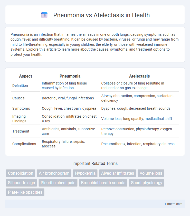
Table of Comparison
| Aspect | Pneumonia | Atelectasis |
|---|---|---|
| Definition | Inflammation of lung tissue caused by infection | Collapse or closure of lung resulting in reduced or no gas exchange |
| Causes | Bacterial, viral, fungal infections | Airway obstruction, compression, surfactant deficiency |
| Symptoms | Cough, fever, chest pain, dyspnea | Dyspnea, cough, decreased breath sounds |
| Imaging Findings | Consolidation, infiltrates on chest X-ray | Volume loss, lung opacity, mediastinal shift |
| Treatment | Antibiotics, antivirals, supportive care | Remove obstruction, physiotherapy, oxygen therapy |
| Complications | Respiratory failure, sepsis, abscess | Pneumothorax, infection, respiratory distress |
Introduction to Pneumonia and Atelectasis
Pneumonia is an inflammatory condition of the lung primarily caused by bacterial, viral, or fungal infections, leading to alveolar consolidation and impaired gas exchange. Atelectasis refers to the partial or complete collapse of lung tissue, resulting in reduced or absent gas exchange in affected areas due to airway obstruction, compression, or surfactant deficiency. Both conditions significantly impact respiratory function but differ in etiology, presentation, and treatment approaches.
Definition of Pneumonia
Pneumonia is an acute respiratory infection characterized by inflammation of the alveoli and lung parenchyma, typically caused by bacterial, viral, or fungal pathogens. It leads to alveolar filling with pus or fluid, impairing gas exchange and causing symptoms like cough, fever, and difficulty breathing. Pneumonia differs from atelectasis, which is the collapse or incomplete expansion of lung tissue without infectious inflammation.
Definition of Atelectasis
Atelectasis is the partial or complete collapse of a lung or a section (lobe) of a lung, resulting in reduced or absent gas exchange in the affected areas. It occurs when alveoli, the tiny air sacs in the lungs, become deflated or filled with fluid, leading to impaired oxygen delivery to the bloodstream. Unlike pneumonia, which is an infection causing inflammation and fluid buildup in the lungs, atelectasis primarily involves physical collapse and can result from airway obstruction, compression, or post-surgical complications.
Key Differences Between Pneumonia and Atelectasis
Pneumonia is an infection causing inflammation and fluid buildup in the alveoli, while atelectasis involves the collapse of lung tissue leading to reduced air volume. Pneumonia typically presents with fever, productive cough, and infiltrates visible on chest X-rays, whereas atelectasis shows lung volume loss and shifted structures without infectious symptoms. Treatment for pneumonia requires antibiotics targeting the causative pathogen, but atelectasis management focuses on re-expanding the lung through respiratory therapy or addressing underlying obstructions.
Common Causes of Pneumonia
Pneumonia commonly results from bacterial infections such as Streptococcus pneumoniae, Haemophilus influenzae, and Mycoplasma pneumoniae, as well as viral pathogens like influenza and respiratory syncytial virus. Risk factors include aspiration due to impaired swallowing, chronic lung diseases, smoking, and immunosuppression. Differentiating pneumonia from atelectasis is critical as pneumonia involves infection and inflammation of the lung parenchyma, whereas atelectasis primarily refers to lung collapse or incomplete expansion.
Common Causes of Atelectasis
Atelectasis commonly results from airway obstruction caused by mucus plugs, tumors, or foreign bodies, leading to collapsed lung tissue and impaired gas exchange. Postoperative complications, prolonged bed rest, and shallow breathing also contribute to atelectasis by preventing full lung expansion. Unlike pneumonia, which typically arises from infections like bacteria or viruses, atelectasis primarily involves mechanical factors obstructing airflow or reducing lung ventilation.
Signs and Symptoms Comparison
Pneumonia commonly presents with fever, productive cough, dyspnea, and pleuritic chest pain, often accompanied by crackles and bronchial breath sounds on auscultation. Atelectasis primarily manifests as dyspnea, tachypnea, decreased breath sounds, and dullness to percussion over the affected area, frequently without fever or significant cough. Both conditions can cause hypoxemia and respiratory distress, but pneumonia typically involves systemic signs of infection absent in atelectasis.
Diagnosis: Pneumonia vs Atelectasis
Diagnosis of pneumonia versus atelectasis relies heavily on imaging techniques such as chest X-rays and CT scans, where pneumonia typically presents with localized alveolar consolidation and air bronchograms, whereas atelectasis shows volume loss with displacement of surrounding structures. Laboratory tests, including elevated inflammatory markers and positive sputum cultures, support a pneumonia diagnosis but are usually absent in atelectasis. Clinical evaluation emphasizing fever, productive cough, and leukocytosis favors pneumonia, while dyspnea and hypoxemia without infection signs suggest atelectasis.
Treatment Approaches for Each Condition
Pneumonia treatment primarily involves targeted antibiotic therapy based on the suspected pathogen, supportive care with oxygen supplementation, and in severe cases, hospitalization for intravenous antibiotics and respiratory support. Atelectasis management focuses on relieving airway obstruction through chest physiotherapy, incentive spirometry, and addressing underlying causes such as mucus plugs or tumors, often complemented by bronchoscopy for mucus clearance. Both conditions require monitoring for complications, but pneumonia demands more aggressive antimicrobial strategies, whereas atelectasis emphasizes mechanical re-expansion of collapsed lung tissue.
Prevention and Risk Reduction Strategies
Preventing pneumonia and atelectasis involves maintaining strong respiratory health through vaccination, smoking cessation, and regular hand hygiene to reduce infection risks. Early mobilization and deep breathing exercises post-surgery or during illness help minimize atelectasis by promoting lung expansion and secretion clearance. Managing underlying conditions such as chronic obstructive pulmonary disease (COPD) and ensuring adequate hydration further reduce the likelihood of both respiratory complications.
Pneumonia Infographic