Hypoplasia is a developmental condition characterized by the underdevelopment or incomplete formation of tissues or organs, resulting in functional limitations. It commonly affects bones, teeth, and various organs, potentially leading to complications depending on the severity and location. Explore this article to understand hypoplasia's causes, symptoms, and treatment options to better manage your health concerns.
Table of Comparison
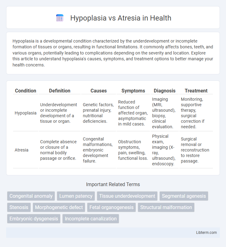
| Condition | Definition | Causes | Symptoms | Diagnosis | Treatment |
|---|---|---|---|---|---|
| Hypoplasia | Underdevelopment or incomplete development of a tissue or organ. | Genetic factors, prenatal injury, nutritional deficiencies. | Reduced function of affected organ, asymptomatic in mild cases. | Imaging (MRI, ultrasound), biopsy, clinical evaluation. | Monitoring, supportive therapy, surgical correction if needed. |
| Atresia | Complete absence or closure of a normal bodily passage or orifice. | Congenital malformations, embryonic development failure. | Obstruction symptoms, pain, swelling, functional loss. | Physical exam, imaging (X-ray, ultrasound), endoscopy. | Surgical removal or reconstruction to restore passage. |
Understanding Hypoplasia: Overview and Definition
Hypoplasia refers to the underdevelopment or incomplete formation of a tissue or organ, resulting in a smaller size and reduced function compared to normal. It is a developmental anomaly characterized by a decreased number of cells, often identified in organs such as the lungs, kidneys, or bones. In contrast, atresia involves the absence or closure of a normal body opening or tubular structure, leading to complete obstruction.
Defining Atresia: Key Concepts
Atresia refers to the complete absence or abnormal closure of a bodily passage or orifice, often congenital in nature, resulting in obstruction of normal physiological flow. Key concepts include its manifestation in various organs such as the esophagus, anus, or bile ducts, and its classification based on the location and severity of the closure. Differentiating atresia from hypoplasia, which involves underdevelopment rather than absence, is critical for accurate diagnosis and treatment planning.
Etiology: What Causes Hypoplasia and Atresia?
Hypoplasia results from incomplete or underdeveloped tissue growth during fetal development, often caused by genetic mutations, chromosomal abnormalities, or environmental factors such as maternal infections and nutritional deficiencies. Atresia occurs due to the failure of a normal body passage or lumen to develop or canalize, commonly linked to vascular disruptions, teratogenic exposures, or congenital malformations affecting organogenesis. Both conditions reflect developmental anomalies but differ fundamentally in their embryological origins and pathological presentations.
Embryological Differences: Hypoplasia vs Atresia
Hypoplasia results from incomplete or underdeveloped organ formation during embryogenesis, characterized by reduced cell proliferation and differentiation, whereas atresia involves the complete absence or closure of a normal body orifice or tubular organ due to failed canalization or recanalization processes in the embryo. These defects arise from distinct disruptions in developmental pathways, with hypoplasia linked to impaired growth signaling and atresia associated with abnormal tissue morphogenesis or apoptosis. Understanding embryological timing and mechanism differences is crucial for accurate diagnosis and targeted therapeutic strategies.
Clinical Manifestations: How Symptoms Differ
Hypoplasia presents with underdeveloped organs or tissues, causing mild to moderate functional impairment such as reduced size or partial blockage in affected areas, often leading to symptoms like fatigue or localized discomfort. Atresia involves a complete absence or closure of a body canal or passage, resulting in severe symptoms like obstruction, absence of normal function, and urgent clinical signs depending on the organ involved. Differentiating these conditions relies on symptom severity and diagnostic imaging, with hypoplasia causing less acute symptoms compared to the often life-threatening manifestations of atresia.
Diagnostic Approaches: Identifying Hypoplasia and Atresia
Diagnostic approaches for hypoplasia and atresia primarily involve advanced imaging techniques such as ultrasound, MRI, and CT scans to assess the structural development and patency of organs or vessels. Hypoplasia is identified by underdeveloped but present anatomical structures showing reduced size or incomplete formation, whereas atresia is characterized by the complete absence or closure of a normal bodily passage. Definitive diagnosis often requires combining imaging findings with clinical evaluation, biopsy, or surgical exploration to differentiate between these congenital anomalies accurately.
Common Examples and Affected Organs
Hypoplasia commonly affects organs like the lungs, kidneys, and heart, characterized by underdeveloped tissue leading to reduced functionality, with examples including renal hypoplasia and pulmonary hypoplasia. Atresia involves the complete absence or closure of a normal body opening or tubular structure, frequently seen in conditions such as esophageal atresia and biliary atresia. Both conditions impact critical organs but differ in severity, with hypoplasia resulting in incomplete development and atresia causing total obstruction or absence.
Treatment Strategies: Tailored Therapies
Hypoplasia treatment strategies prioritize organ function enhancement through progressive therapies such as physical rehabilitation or growth stimulation, whereas atresia often requires surgical intervention to restore anatomical continuity, such as anastomosis or stenting. Personalized treatment plans incorporate diagnostic imaging and genetic testing to tailor therapies based on severity and affected areas, optimizing patient outcomes. Emerging approaches include regenerative medicine and minimally invasive surgeries to reduce complications and improve recovery times in both conditions.
Prognosis and Long-Term Outcomes
Hypoplasia involves underdevelopment of an organ or tissue, leading to variable prognosis depending on severity and timely intervention, while atresia is a complete absence or closure of a normal body opening or tubular structure, often requiring immediate surgical correction. Long-term outcomes in hypoplasia can range from mild functional impairment to significant disability, whereas atresia often demands lifelong management and can result in chronic complications like infections or impaired organ function. Early diagnosis and specialized treatment improve prognosis in both conditions but emphasize the critical nature of early intervention in atresia for survival and quality of life.
Key Differences: Hypoplasia vs Atresia Compared
Hypoplasia refers to the underdevelopment or incomplete development of a tissue or organ, resulting in a smaller size but retaining some functional capacity, whereas atresia involves the complete absence or closure of a normal body opening or tubular structure, leading to obstruction. Hypoplasia often presents with reduced cell number and size but maintains some degree of structure, while atresia is characterized by a congenital absence or sealed lumen, causing functional impairment. The critical difference lies in hypoplasia's partial organ formation versus atresia's total discontinuity or obstruction of the anatomical passage.
Hypoplasia Infographic