Hyperplastic conditions involve excessive cell proliferation leading to tissue enlargement or abnormal growths, often signaling an underlying pathological process. Understanding the causes and symptoms of hyperplastic changes can help in early diagnosis and effective management of related disorders. Explore the rest of the article to learn how hyperplastic developments may impact your health and treatment options.
Table of Comparison
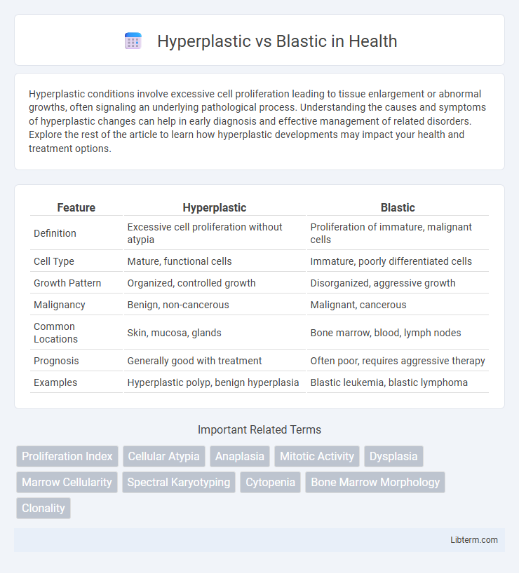
| Feature | Hyperplastic | Blastic |
|---|---|---|
| Definition | Excessive cell proliferation without atypia | Proliferation of immature, malignant cells |
| Cell Type | Mature, functional cells | Immature, poorly differentiated cells |
| Growth Pattern | Organized, controlled growth | Disorganized, aggressive growth |
| Malignancy | Benign, non-cancerous | Malignant, cancerous |
| Common Locations | Skin, mucosa, glands | Bone marrow, blood, lymph nodes |
| Prognosis | Generally good with treatment | Often poor, requires aggressive therapy |
| Examples | Hyperplastic polyp, benign hyperplasia | Blastic leukemia, blastic lymphoma |
Introduction to Hyperplastic and Blastic Processes
Hyperplastic processes involve an increase in the number of cells, leading to tissue enlargement without altering the cellular architecture, commonly seen in conditions like benign prostatic hyperplasia. Blastic processes, typified by excessive formation of immature cells, often occur in aggressive malignancies such as blastic leukemia, characterized by rapid proliferation and impaired differentiation. Understanding the distinction between hyperplastic and blastic tissue growth is crucial for accurate diagnosis and therapeutic strategy formulation in pathological conditions.
Defining Hyperplastic: Characteristics and Mechanisms
Hyperplastic refers to the increased proliferation of normal cells resulting in tissue enlargement without malignant transformation, characterized by an increased cell number maintaining normal architecture and function. Mechanisms involve stimuli triggering cellular mitotic activity, such as hormonal signals or chronic irritation, leading to reversible tissue growth. Unlike blastic processes, which often imply abnormal, neoplastic bone formation, hyperplastic changes are controlled and adaptive responses maintaining tissue homeostasis.
Understanding Blastic: Key Features and Pathways
Blastic lesions are characterized by increased bone formation due to enhanced osteoblastic activity, often resulting in sclerotic or dense radiographic appearances. Key molecular pathways involved in blastic bone formation include the Wnt/b-catenin signaling and bone morphogenetic proteins (BMPs), which stimulate osteoblast differentiation and matrix mineralization. Understanding these signaling cascades is critical for developing targeted therapies in diseases such as blastic bone metastases and osteosclerotic conditions.
Cellular Differences: Hyperplastic vs Blastic
Hyperplastic tissue displays an increased number of normal cells due to heightened cell proliferation, maintaining typical cellular morphology and function. Blastic tissue, however, features immature, undifferentiated cells with high mitotic activity and abnormal nuclei, indicating a pathological or neoplastic process. The key cellular difference lies in hyperplastic cells retaining normal differentiation, whereas blastic cells exhibit primitive, aberrant characteristics often linked to malignancy.
Clinical Implications of Hyperplastic Conditions
Hyperplastic conditions involve an increase in the number of cells, often leading to tissue enlargement without atypia or malignancy risk, which generally suggests a benign clinical course. These conditions, such as benign prostatic hyperplasia or reactive lymphoid hyperplasia, require monitoring for symptomatic management rather than aggressive treatment. Distinguishing hyperplastic lesions from blastic (malignant or neoplastic) conditions is crucial for prognosis and therapeutic decisions, highlighting the importance of accurate histopathological evaluation.
Blastic Lesions: Clinical Significance
Blastic lesions, characterized by increased bone formation and density, often indicate aggressive pathological processes such as metastatic prostate cancer or osteosarcoma. Their clinical significance lies in their potential to cause pain, structural bone weakness, and functional impairment, requiring prompt diagnosis and intervention. Advanced imaging techniques and biopsy are essential for differentiating blastic lesions from hyperplastic or sclerotic bone conditions, guiding effective treatment strategies.
Diagnostic Approaches: Differentiating Hyperplastic from Blastic
Differentiating hyperplastic from blastic lesions primarily relies on histopathological examination, where hyperplastic tissues show increased cellular proliferation without malignant features, while blastic lesions demonstrate abnormal, immature cell growth indicative of neoplasia. Imaging techniques such as MRI and CT scans aid in identifying lesion characteristics, with blastic lesions often appearing more aggressive and infiltrative compared to hyperplastic changes. Immunohistochemical markers and molecular diagnostics further enhance accuracy by highlighting specific protein expressions unique to blastic pathology, enabling precise diagnosis and targeted treatment planning.
Common Diseases Associated with Hyperplastic Changes
Hyperplastic changes frequently occur in benign prostatic hyperplasia (BPH), where stromal and epithelial cell proliferation causes prostate enlargement. Hyperplasia is also common in endometrial hyperplasia, often linked to estrogen imbalance and a risk factor for endometrial carcinoma. In bone marrow, hyperplastic marrow is typically reactive, seen in anemia or chronic inflammatory conditions, contrasting with blastic proliferations in malignancies like leukemia.
Disorders Linked to Blastic Activity
Blastic activity is primarily associated with disorders characterized by excessive bone formation, such as osteosclerosis and certain types of bone tumors like osteosarcoma. These conditions often involve abnormal proliferation of osteoblasts leading to increased bone density and structural abnormalities. Understanding blastic activity is crucial for diagnosing and managing diseases that result in pathological bone growth and remodeling.
Prognosis and Treatment Strategies: Hyperplastic vs Blastic
Hyperplastic lesions generally have a better prognosis due to their benign nature, often responding well to conservative treatments such as surgical excision and close monitoring. Blastic lesions, associated with aggressive or malignant growth, typically require more intensive treatment strategies including chemotherapy, radiation therapy, or targeted therapies to improve patient outcomes. Prognostic factors vary significantly between hyperplastic and blastic types, with blastic lesions demonstrating higher recurrence rates and poorer long-term survival.
Hyperplastic Infographic