Gallstones form when substances in bile, such as cholesterol or bilirubin, concentrate and crystallize, leading to cholelithiasis. This condition can cause pain, nausea, and digestive issues if stones block bile ducts. Discover the symptoms, causes, and treatment options to better understand how cholelithiasis may impact your health.
Table of Comparison
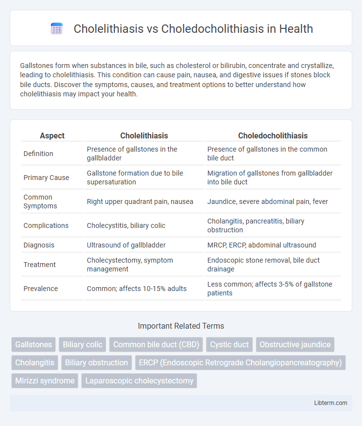
| Aspect | Cholelithiasis | Choledocholithiasis |
|---|---|---|
| Definition | Presence of gallstones in the gallbladder | Presence of gallstones in the common bile duct |
| Primary Cause | Gallstone formation due to bile supersaturation | Migration of gallstones from gallbladder into bile duct |
| Common Symptoms | Right upper quadrant pain, nausea | Jaundice, severe abdominal pain, fever |
| Complications | Cholecystitis, biliary colic | Cholangitis, pancreatitis, biliary obstruction |
| Diagnosis | Ultrasound of gallbladder | MRCP, ERCP, abdominal ultrasound |
| Treatment | Cholecystectomy, symptom management | Endoscopic stone removal, bile duct drainage |
| Prevalence | Common; affects 10-15% adults | Less common; affects 3-5% of gallstone patients |
Overview of Cholelithiasis and Choledocholithiasis
Cholelithiasis refers to the presence of gallstones within the gallbladder, commonly caused by cholesterol supersaturation or pigment stone formation, leading to biliary colic or asymptomatic presentations. Choledocholithiasis involves gallstones lodged in the common bile duct, frequently resulting in obstructive jaundice, cholangitis, or pancreatitis due to impaired bile flow. Diagnosis relies on imaging techniques such as ultrasound for cholelithiasis and magnetic resonance cholangiopancreatography (MRCP) or endoscopic retrograde cholangiopancreatography (ERCP) for choledocholithiasis.
Definition: Cholelithiasis vs Choledocholithiasis
Cholelithiasis refers to the presence of gallstones primarily within the gallbladder, leading to potential biliary obstruction or inflammation. Choledocholithiasis describes gallstones that have migrated into the common bile duct, causing bile duct obstruction and increasing the risk of cholangitis or pancreatitis. Both conditions involve gallstone formation but differ in anatomical location and clinical implications.
Etiology and Risk Factors
Cholelithiasis involves the formation of gallstones primarily in the gallbladder, caused by cholesterol supersaturation, bile stasis, and imbalance in bile salts, influenced by risk factors such as obesity, female gender, pregnancy, and rapid weight loss. Choledocholithiasis refers to gallstones located in the common bile duct, often originating from migrating gallstones or secondary pigment stones formed due to hemolytic disorders or biliary infections. Both conditions share risk factors like advancing age, cholecystitis history, and certain metabolic diseases, but choledocholithiasis is more associated with biliary obstruction and related complications.
Clinical Presentation and Symptoms
Cholelithiasis typically presents with episodic right upper quadrant pain, often postprandial, accompanied by nausea and vomiting without systemic signs. Choledocholithiasis manifests more severely with persistent right upper quadrant pain, jaundice, dark urine, pale stools, and signs of cholangitis such as fever and chills. Laboratory findings in choledocholithiasis include elevated bilirubin, alkaline phosphatase, and transaminases, reflecting biliary obstruction and inflammation.
Diagnostic Approaches for Each Condition
Cholelithiasis is primarily diagnosed through abdominal ultrasound, which effectively detects gallstones within the gallbladder, while liver function tests help assess potential biliary obstruction. Choledocholithiasis requires more advanced imaging such as magnetic resonance cholangiopancreatography (MRCP) or endoscopic retrograde cholangiopancreatography (ERCP) to visualize stones within the common bile duct and evaluate ductal obstruction. Both conditions may involve elevated bilirubin and alkaline phosphatase levels, but choledocholithiasis often presents with more pronounced cholestatic enzyme abnormalities necessitating targeted diagnostic interventions.
Imaging Modalities: Ultrasound, CT, and MRCP
Ultrasound is the primary imaging modality for diagnosing cholelithiasis, effectively detecting gallstones within the gallbladder with high sensitivity and specificity. CT scans provide detailed cross-sectional images but are less sensitive for gallstones compared to ultrasound, though they are valuable in identifying complications like inflammation or biliary obstruction. Magnetic Resonance Cholangiopancreatography (MRCP) excels in visualizing the biliary tree, making it the gold standard for detecting choledocholithiasis by noninvasively identifying stones within the common bile duct.
Complications and Associated Diseases
Cholelithiasis, characterized by gallstone formation in the gallbladder, often leads to complications such as acute cholecystitis, gallbladder empyema, and biliary colic. Choledocholithiasis, involving gallstones in the common bile duct, frequently causes biliary obstruction, cholangitis, and pancreatitis. Both conditions are associated with diseases like diabetes mellitus, obesity, and hemolytic disorders, which increase the risk of gallstone formation and subsequent complications.
Treatment Options: Medical and Surgical
Cholelithiasis treatment often involves watchful waiting for asymptomatic cases, while symptomatic patients may require laparoscopic cholecystectomy to remove gallstones. Choledocholithiasis typically necessitates endoscopic retrograde cholangiopancreatography (ERCP) for stone extraction, followed by cholecystectomy to prevent recurrence. In complicated cases with infections or biliary obstruction, antibiotics and biliary drainage may be essential components of the treatment strategy.
Prognosis and Long-Term Management
Cholelithiasis, characterized by the presence of gallstones within the gallbladder, generally has a favorable prognosis with low complication rates when asymptomatic, while symptomatic cases may require lithotripsy or cholecystectomy to prevent recurrent biliary colic or cholecystitis. Choledocholithiasis involves gallstones in the common bile duct and carries a higher risk of complications like cholangitis or pancreatitis, necessitating prompt endoscopic retrograde cholangiopancreatography (ERCP) for stone extraction to reduce morbidity and mortality. Long-term management of choledocholithiasis often involves monitoring for bile duct strictures or recurrent stones, with prophylactic antibiotics and regular imaging follow-ups, whereas cholelithiasis patients post-cholecystectomy typically require minimal follow-up unless complications occur.
Prevention and Patient Education
Preventing cholelithiasis and choledocholithiasis involves maintaining a healthy weight through a balanced diet rich in fiber and low in saturated fats, which reduces gallstone formation risk. Patient education should emphasize recognizing symptoms early, such as biliary colic and jaundice, to prompt timely medical evaluation and avoid complications like cholangitis or pancreatitis. Regular follow-ups and lifestyle modifications, including hydration and physical activity, are critical to minimizing recurrence and ensuring gallbladder health.
Cholelithiasis Infographic