Empyema is a severe infection characterized by the accumulation of pus in the pleural space, often resulting from untreated pleural effusion or pneumonia. Pleural effusion, the buildup of excess fluid between the layers of the pleura surrounding the lungs, can lead to breathing difficulties and requires timely diagnosis and intervention. Explore this article to understand the causes, symptoms, and treatment options that could impact your lung health.
Table of Comparison
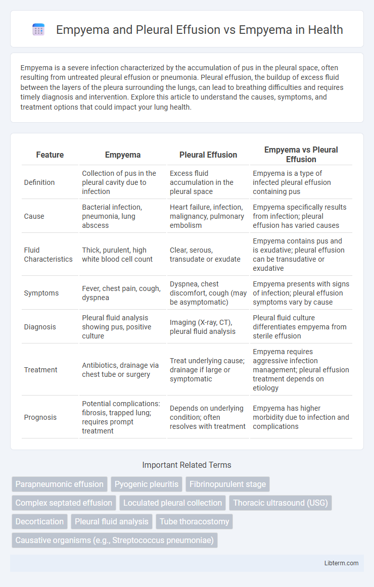
| Feature | Empyema | Pleural Effusion | Empyema vs Pleural Effusion |
|---|---|---|---|
| Definition | Collection of pus in the pleural cavity due to infection | Excess fluid accumulation in the pleural space | Empyema is a type of infected pleural effusion containing pus |
| Cause | Bacterial infection, pneumonia, lung abscess | Heart failure, infection, malignancy, pulmonary embolism | Empyema specifically results from infection; pleural effusion has varied causes |
| Fluid Characteristics | Thick, purulent, high white blood cell count | Clear, serous, transudate or exudate | Empyema contains pus and is exudative; pleural effusion can be transudative or exudative |
| Symptoms | Fever, chest pain, cough, dyspnea | Dyspnea, chest discomfort, cough (may be asymptomatic) | Empyema presents with signs of infection; pleural effusion symptoms vary by cause |
| Diagnosis | Pleural fluid analysis showing pus, positive culture | Imaging (X-ray, CT), pleural fluid analysis | Pleural fluid culture differentiates empyema from sterile effusion |
| Treatment | Antibiotics, drainage via chest tube or surgery | Treat underlying cause; drainage if large or symptomatic | Empyema requires aggressive infection management; pleural effusion treatment depends on etiology |
| Prognosis | Potential complications: fibrosis, trapped lung; requires prompt treatment | Depends on underlying condition; often resolves with treatment | Empyema has higher morbidity due to infection and complications |
Understanding Pleural Effusion: Definition and Causes
Pleural effusion is the abnormal accumulation of fluid in the pleural space, caused by conditions such as heart failure, pneumonia, malignancy, or pulmonary embolism. Empyema is a type of pleural effusion characterized by the presence of pus, indicating an infection within the pleural cavity. Understanding pleural effusion involves recognizing that its causes range from transudative fluid due to systemic factors to exudative fluid resulting from local inflammation or infection, as seen in empyema.
What Is Empyema? Understanding the Distinction
Empyema is a collection of pus within the pleural cavity, often caused by bacterial infections leading to an inflammatory response and lung tissue damage. Pleural effusion involves the accumulation of excess fluid between the layers of the pleura but can be either transudative or exudative, with empyema representing a specific type of infected, purulent pleural effusion. Differentiating empyema from pleural effusion is crucial for treatment, as empyema requires prompt drainage and antibiotics to prevent complications like fibrosis and sepsis.
Pathophysiology: Pleural Effusion vs Empyema
Pleural effusion involves the accumulation of excess fluid in the pleural space due to imbalance in hydrostatic and oncotic pressures or increased capillary permeability, often from heart failure, infection, or malignancy. Empyema represents a progression of pleural effusion where the fluid becomes infected and purulent, leading to fibrin deposition, pleural thickening, and impaired lung expansion. The pathophysiological distinction lies in the shift from sterile fluid accumulation in pleural effusion to bacterial invasion and pus formation in empyema, triggering an intense inflammatory response and fibrosis.
Clinical Presentation: Similarities and Differences
Empyema and pleural effusion both present with symptoms such as chest pain, fever, and dyspnea due to fluid accumulation in the pleural space. Empyema, a purulent pleural effusion caused by infection, typically shows more pronounced systemic signs like high-grade fever and chills compared to sterile pleural effusion. Clinical differentiation relies on thoracentesis revealing pus and positive bacterial cultures in empyema, whereas pleural effusion fluid is often clear or serous with negative cultures.
Diagnostic Modalities for Pleural Diseases
Diagnostic modalities for pleural diseases such as pleural effusion and empyema primarily include chest X-rays, which identify fluid accumulation and lung compression, and ultrasound, which effectively differentiates between simple pleural effusion and complex empyema by detecting septations and loculations. Computed tomography (CT) scans provide detailed imaging to assess pleural thickening, lung entrapment, and guide thoracentesis or chest tube placement. Pleural fluid analysis through thoracentesis remains essential for distinguishing empyema, characterized by purulent fluid and positive microbiological cultures, from non-infectious pleural effusions.
Imaging Features: Differentiating Empyema from Pleural Effusion
Imaging features play a crucial role in differentiating empyema from pleural effusion, with ultrasound and CT being the primary modalities. Empyema typically presents as complex, septated fluid collections with thick, enhancing pleural walls on contrast-enhanced CT, whereas simple pleural effusions appear as homogenous, non-septated anechoic or hypoechoic fluid without pleural thickening. The presence of loculations, internal debris, and pleural enhancement strongly suggests empyema, distinguishing it from uncomplicated pleural effusions that lack these features.
Laboratory Analysis: Thoracentesis Findings
Thoracentesis in pleural effusion typically reveals clear, straw-colored fluid with low white blood cell count, predominantly lymphocytes or macrophages, and a protein level that helps classify the effusion as transudate or exudate. In empyema, thoracentesis fluid is purulent, with high neutrophil count, elevated lactate dehydrogenase (LDH), low glucose levels, and positive bacterial cultures indicating infection. Laboratory analysis of thoracentesis fluid differentiates empyema from simple pleural effusion by its characteristic biochemical markers and microbial presence.
Treatment Approaches: Pleural Effusion vs Empyema
Treatment approaches for pleural effusion primarily involve thoracentesis to drain excess fluid and addressing the underlying cause, such as infection or malignancy. Empyema treatment requires more aggressive interventions including chest tube drainage combined with prolonged antibiotic therapy to eradicate pus-filled infected pleural fluid. Surgical options like video-assisted thoracoscopic surgery (VATS) or decortication are often necessary in empyema cases to remove fibrinous peel and facilitate lung re-expansion.
Prognosis and Potential Complications
Empyema, characterized by pus accumulation in the pleural space, generally has a more severe prognosis than simple pleural effusion due to its infectious nature and risk of progressing to fibrothorax or sepsis. Pleural effusion, often caused by heart failure, malignancy, or infection, presents varied outcomes depending on underlying etiology, but complications are less extensive unless complicated by infection leading to empyema. Early diagnosis and effective drainage, combined with appropriate antibiotic therapy, are critical in improving prognosis and preventing complications such as lung entrapment or chronic pleural thickening in empyema cases.
Prevention and Early Intervention Strategies
Empyema, a collection of pus in the pleural space usually caused by bacterial infection, differs from simple pleural effusion, which is the accumulation of excess fluid without infection. Prevention strategies for empyema emphasize prompt treatment of pneumonia and respiratory infections, along with vaccination against Streptococcus pneumoniae and influenza to reduce incidence. Early intervention involves rapid diagnostic imaging and thoracentesis to drain infected fluid, combined with appropriate antibiotic therapy to prevent progression and complications.
Empyema and Pleural Effusion Infographic