Lingual hemangioma is a benign vascular tumor occurring on the tongue, often appearing as a red or purple raised lesion due to an abnormal buildup of blood vessels. These hemangiomas can cause discomfort, bleeding, and difficulties with speech or swallowing depending on their size and location. Explore the rest of the article to understand symptoms, treatment options, and care for your lingual hemangioma.
Table of Comparison
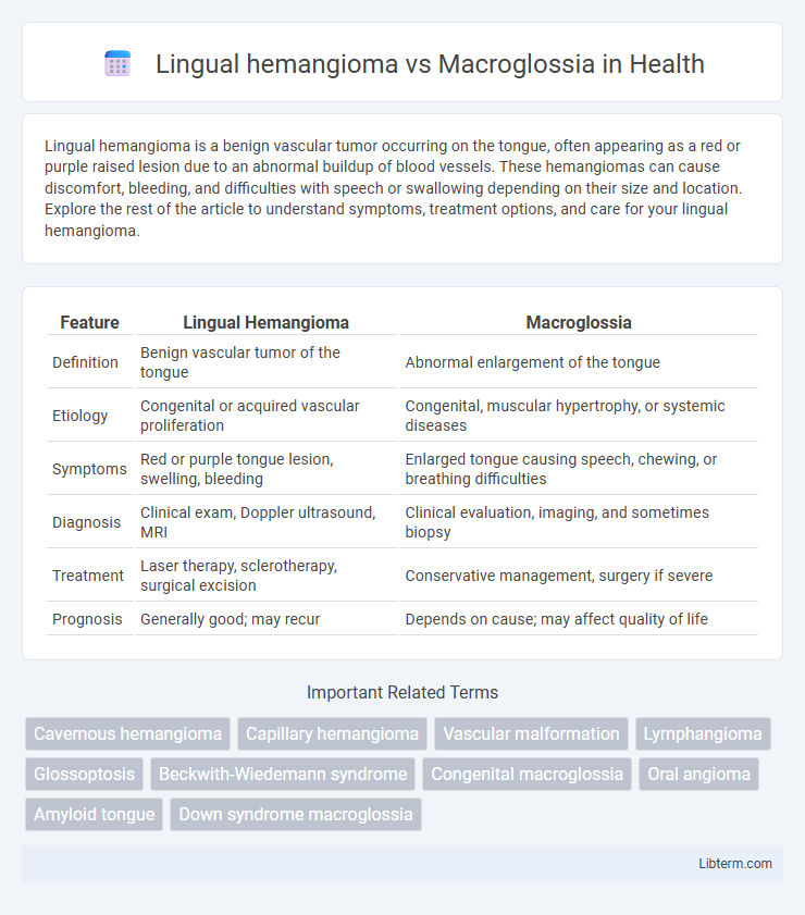
| Feature | Lingual Hemangioma | Macroglossia |
|---|---|---|
| Definition | Benign vascular tumor of the tongue | Abnormal enlargement of the tongue |
| Etiology | Congenital or acquired vascular proliferation | Congenital, muscular hypertrophy, or systemic diseases |
| Symptoms | Red or purple tongue lesion, swelling, bleeding | Enlarged tongue causing speech, chewing, or breathing difficulties |
| Diagnosis | Clinical exam, Doppler ultrasound, MRI | Clinical evaluation, imaging, and sometimes biopsy |
| Treatment | Laser therapy, sclerotherapy, surgical excision | Conservative management, surgery if severe |
| Prognosis | Generally good; may recur | Depends on cause; may affect quality of life |
Introduction to Lingual Hemangioma and Macroglossia
Lingual hemangioma is a benign vascular tumor commonly found on the tongue, characterized by the proliferation of blood vessels leading to a reddish or purplish lesion. Macroglossia refers to an abnormal enlargement of the tongue, often caused by underlying conditions such as congenital anomalies, muscular hypertrophy, or vascular malformations including hemangiomas. Understanding the distinct clinical presentations and etiologies of lingual hemangioma and macroglossia is essential for accurate diagnosis and effective treatment planning.
Definition and Clinical Features
Lingual hemangioma is a benign vascular tumor characterized by a proliferation of blood vessels primarily affecting the tongue, presenting clinically as a red to purple, soft, compressible mass that may cause bleeding or ulceration. Macroglossia refers to an abnormal enlargement of the tongue due to muscular hypertrophy, infiltration by abnormal tissue, or vascular abnormalities, often leading to difficulties in speech, swallowing, and airway obstruction. Both conditions can present with tongue swelling, but lingual hemangioma is distinguished by its vascular nature and potential for spontaneous regression, whereas macroglossia is generally a structural enlargement with a more persistent course.
Etiology and Pathogenesis
Lingual hemangioma is a benign vascular tumor resulting from endothelial cell proliferation, often linked to somatic mutations in the VEGF pathway causing abnormal blood vessel formation. Macroglossia involves tongue enlargement due to diverse causes such as muscular hypertrophy, lymphatic malformations, or systemic conditions like hypothyroidism and amyloidosis, leading to tissue overgrowth or infiltration. While lingual hemangioma primarily arises from localized vascular anomalies, macroglossia reflects broader etiologies associated with structural or metabolic abnormalities affecting tongue size and function.
Epidemiology and Risk Factors
Lingual hemangiomas primarily affect infants, with a higher incidence in females and premature infants, often presenting within the first few weeks of life due to abnormal proliferation of blood vessels. Macroglossia is less common and can be congenital or acquired, frequently associated with conditions such as Down syndrome, Beckwith-Wiedemann syndrome, and hypothyroidism, affecting both pediatric and adult populations. Risk factors for lingual hemangioma include low birth weight and female gender, while macroglossia risk factors are linked to genetic syndromes, metabolic disorders, and vascular malformations.
Diagnostic Criteria and Techniques
Lingual hemangioma is diagnosed through clinical examination and confirmed by imaging techniques such as ultrasound Doppler and MRI, which reveal a well-defined vascular lesion with characteristic blood flow patterns, whereas macroglossia involves an enlarged tongue without vascular anomalies, often evaluated by physical measurement and imaging like MRI or CT to assess tissue hypertrophy and underlying causes. Biopsy and histopathological analysis are critical in distinguishing vascular proliferation in hemangiomas from muscular hypertrophy seen in macroglossia. Differential diagnosis relies heavily on history, lesion consistency, color, and growth behavior, with hemangiomas typically present at birth or early childhood and showing rapid initial growth followed by involution, while macroglossia is commonly congenital or secondary to systemic conditions.
Histopathology and Imaging Findings
Lingual hemangioma presents histopathologically with proliferating endothelial cells forming numerous vascular channels, often filled with erythrocytes, while macroglossia shows muscle hypertrophy or infiltration of abnormal tissue such as lymphangiomatous or muscular hypertrophy changes. Imaging findings in lingual hemangioma typically reveal a well-defined, hypervascular lesion on Doppler ultrasound and intense contrast enhancement on MRI due to blood flow within the vascular spaces. Macroglossia exhibits diffuse tongue enlargement with heterogeneous muscle architecture on MRI, often without the distinct vascular enhancement characteristic of hemangiomas.
Clinical Manifestations and Complications
Lingual hemangioma presents as a red or purple, compressible vascular lesion on the tongue, often causing swelling, bleeding, or ulceration, while macroglossia is characterized by an enlarged tongue due to muscular or vascular hypertrophy, frequently leading to speech difficulties, airway obstruction, and dental malocclusion. Complications of lingual hemangioma include hemorrhage and infection, whereas macroglossia can cause chronic airway compromise, feeding problems, and secondary mandibular deformities. Early diagnosis and differentiation between these conditions are vital for appropriate management and prevention of functional impairments.
Treatment Options and Management Strategies
Lingual hemangioma treatment primarily involves laser therapy, corticosteroid injections, or surgical excision depending on size and symptoms, while beta-blockers like propranolol have shown effectiveness in reducing lesion size. Management of macroglossia focuses on addressing functional impairment or airway obstruction through surgical reduction (glossectomy) and supportive therapies such as orthodontic treatment or speech therapy. Regular monitoring and multidisciplinary care including otolaryngologists, oral surgeons, and speech therapists optimize outcomes for both conditions.
Prognosis and Outcomes
Lingual hemangioma typically exhibits a favorable prognosis with many lesions undergoing spontaneous involution by early childhood, resulting in minimal long-term functional impairment or cosmetic concerns. Macroglossia, often associated with congenital syndromes or systemic conditions, may require surgical intervention to improve airway patency, speech, and feeding, with outcomes dependent on underlying etiology and extent of tongue enlargement. Long-term outcomes for macroglossia vary widely, whereas lingual hemangioma generally resolves with low recurrence risk and excellent functional restoration.
Key Differences Between Lingual Hemangioma and Macroglossia
Lingual hemangioma is a benign vascular tumor characterized by a proliferation of blood vessels on the tongue, often presenting as a red or purple lesion that can bleed easily. Macroglossia involves an abnormal enlargement of the entire tongue, usually due to genetic, muscular, or systemic causes, resulting in functional impairments such as speech difficulties or airway obstruction. Key differences include lingual hemangioma's vascular origin and localized nature versus macroglossia's diffuse tongue enlargement and heterogeneous etiology.
Lingual hemangioma Infographic