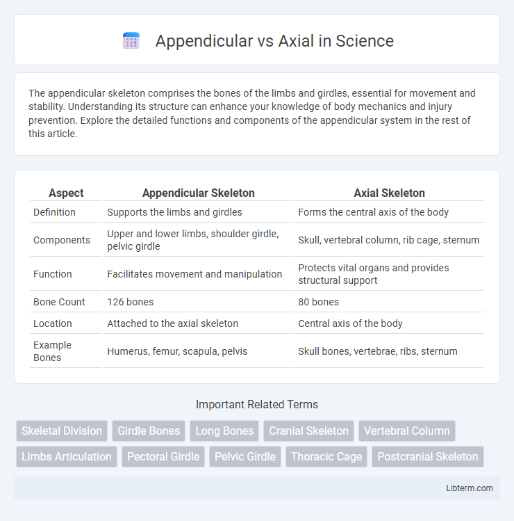
The appendicular skeleton comprises the bones of the limbs and girdles, essential for movement and stability. Understanding its structure can enhance your knowledge of body mechanics and injury prevention. Explore the detailed functions and components of the appendicular system in the rest of this article.
Table of Comparison
| Aspect | Appendicular Skeleton | Axial Skeleton |
|---|---|---|
| Definition | Supports the limbs and girdles | Forms the central axis of the body |
| Components | Upper and lower limbs, shoulder girdle, pelvic girdle | Skull, vertebral column, rib cage, sternum |
| Function | Facilitates movement and manipulation | Protects vital organs and provides structural support |
| Bone Count | 126 bones | 80 bones |
| Location | Attached to the axial skeleton | Central axis of the body |
| Example Bones | Humerus, femur, scapula, pelvis | Skull bones, vertebrae, ribs, sternum |
Introduction to the Skeletal System
The skeletal system is divided into two primary regions: the axial skeleton, consisting of 80 bones including the skull, vertebral column, and rib cage, and the appendicular skeleton, composed of 126 bones in the limbs and girdles. The axial skeleton provides structural support and protection for the brain, spinal cord, and thoracic organs, while the appendicular skeleton facilitates movement and interaction with the environment. Understanding the distinction between these regions is essential for studying human anatomy and biomechanics.
Defining Axial Skeleton
The axial skeleton consists of 80 bones forming the central axis of the body, including the skull, vertebral column, and thoracic cage. It provides structural support, protects the brain, spinal cord, and vital organs, and serves as an attachment site for muscles that facilitate posture and respiration. Unlike the appendicular skeleton, which comprises the limbs and girdles, the axial skeleton is crucial for maintaining the body's stability and protecting its core structures.
Defining Appendicular Skeleton
The appendicular skeleton consists of 126 bones that form the limbs and girdles, facilitating movement and interaction with the environment. It includes the pectoral girdles, upper limbs, pelvic girdle, and lower limbs, supporting mobility and dexterity. This structure contrasts with the axial skeleton, which comprises 80 bones focused on protecting the central nervous system and supporting the body's core.
Major Components of the Axial Skeleton
The axial skeleton consists of 80 bones, including the skull, vertebral column, and thoracic cage, serving as the central framework of the body. Major components such as the cranial bones protect the brain, while the vertebrae support the spinal cord and enable posture. The thoracic cage, composed of ribs and sternum, protects vital organs like the heart and lungs and supports respiration.
Key Structures of the Appendicular Skeleton
The appendicular skeleton comprises 126 bones primarily responsible for movement and includes the key structures of the limbs and girdles. Major components consist of the pectoral girdle (clavicles and scapulae), the upper limbs (humerus, radius, ulna, carpals, metacarpals, and phalanges), the pelvic girdle (ilium, ischium, and pubis), and the lower limbs (femur, patella, tibia, fibula, tarsals, metatarsals, and phalanges). These structures collaborate to facilitate locomotion, manipulation, and support of the body in conjunction with the axial skeleton.
Functional Differences: Axial vs. Appendicular
The axial skeleton provides the central framework for body support and protection of vital organs, including the skull, vertebral column, and rib cage, enabling posture and stability. In contrast, the appendicular skeleton, comprised of limbs and girdles, facilitates movement and interaction with the environment through locomotion and manual dexterity. Functional differences highlight the axial skeleton's role in structural integrity and protection, while the appendicular skeleton is essential for mobility and manipulation of objects.
Developmental Origins and Growth
The appendicular skeleton, consisting of the limbs and girdles, originates primarily from the lateral plate mesoderm, while the axial skeleton, including the skull, vertebral column, and rib cage, develops mainly from the paraxial mesoderm and neural crest cells. During embryogenesis, distinct signaling pathways such as FGF and Wnt regulate the morphogenesis and elongation of appendicular bones, whereas axial skeleton growth relies heavily on the somitic segmentation process and notochordal influences. Postnatal growth of appendicular bones occurs through endochondral ossification at the epiphyseal plates, contrasting with intramembranous ossification in parts of the axial skeleton like the cranial bones.
Common Disorders Affecting Axial and Appendicular Skeletons
Common disorders affecting the axial skeleton include herniated discs, spinal stenosis, and scoliosis, which primarily impact the vertebrae, ribs, and skull. Appendicular skeleton disorders such as osteoarthritis, fractures, and rotator cuff injuries predominantly involve the limbs, shoulder, and pelvic girdles. Both skeletons can also be affected by osteoporosis, leading to increased fracture risk and impaired mobility.
Anatomical Comparison: Axial vs. Appendicular
The axial skeleton consists of 80 bones, including the skull, vertebral column, and rib cage, providing central support and protection for vital organs. The appendicular skeleton comprises 126 bones, encompassing the limbs and girdles, enabling movement and manipulation of the environment. Anatomically, the axial skeleton forms the body's vertical axis, whereas the appendicular skeleton facilitates locomotion and interaction through its attachment to the axial framework.
Clinical Importance and Applications
The axial skeleton, comprising the skull, vertebral column, and rib cage, provides critical protection for vital organs such as the brain, heart, and lungs, making it essential in trauma assessments and neurosurgical planning. The appendicular skeleton, including the limbs and girdles, is key to evaluating mobility impairments and planning orthopedic surgeries like joint replacements or fracture repairs. Understanding the distinctions between these skeletal divisions aids in accurate diagnosis of musculoskeletal disorders and targeted rehabilitation strategies.
Appendicular Infographic

 libterm.com