Atrophy is the gradual decline or shrinkage of tissues, muscles, or organs due to disuse, disease, or malnutrition. This condition can significantly impair physical function and overall health if left unaddressed. Discover how you can prevent and manage atrophy effectively by exploring the rest of this article.
Table of Comparison
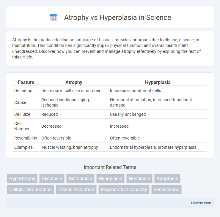
| Feature | Atrophy | Hyperplasia |
|---|---|---|
| Definition | Decrease in cell size or number | Increase in number of cells |
| Cause | Reduced workload, aging, ischemia | Hormonal stimulation, increased functional demand |
| Cell Size | Reduced | Usually unchanged |
| Cell Number | Decreased | Increased |
| Reversibility | Often reversible | Often reversible |
| Examples | Muscle wasting, brain atrophy | Endometrial hyperplasia, prostate hyperplasia |
Introduction to Atrophy and Hyperplasia
Atrophy refers to the reduction in size or number of cells in an organ or tissue, resulting in decreased functionality, often caused by factors such as aging, decreased workload, or nutrient deprivation. Hyperplasia involves an increase in the number of cells in a tissue or organ, leading to enlargement due to stimuli like hormonal changes or chronic irritation. Both atrophy and hyperplasia represent adaptive cellular responses to environmental changes, critical in maintaining homeostasis or signaling pathological conditions.
Defining Atrophy
Atrophy refers to the reduction in size or number of cells in a tissue or organ, leading to a decrease in overall mass and function, often caused by factors such as disuse, malnutrition, aging, or decreased blood supply. This cellular shrinkage contrasts with hyperplasia, which involves an increase in the number of cells, resulting in tissue enlargement. Understanding atrophy is crucial for diagnosing conditions like muscle wasting, brain degeneration, and organ shrinkage in various pathological states.
Defining Hyperplasia
Hyperplasia is the increase in the number of cells in a tissue or organ, leading to its enlargement, often as a response to a stimulus or increased functional demand. Unlike atrophy, which involves a reduction in cell size or number, hyperplasia results from cellular proliferation and can occur physiologically (e.g., during wound healing) or pathologically (e.g., in benign prostatic hyperplasia). The process is tightly regulated by growth factors and hormones to ensure controlled tissue growth and adaptation.
Key Differences Between Atrophy and Hyperplasia
Atrophy involves the reduction in cell size or number resulting in decreased tissue or organ mass, often due to factors like aging, disuse, or nutrient deficiency. Hyperplasia is characterized by an increase in the number of cells, leading to tissue or organ enlargement, typically driven by hormonal signals or compensatory mechanisms. Unlike atrophy, hyperplasia maintains normal cell size and is a regulated proliferative response, whereas atrophy reflects degeneration or a catabolic state.
Causes of Atrophy
Atrophy results from factors that reduce cellular size or number, including diminished blood supply, decreased hormonal stimulation, malnutrition, and prolonged disuse of muscles or organs. Chronic inflammation, aging, and nerve damage contribute to cellular shrinkage and organ regression, distinguishing atrophy from hyperplasia, which involves increased cell proliferation. Understanding these causes is vital for diagnosing diseases related to tissue degeneration and impaired function.
Causes of Hyperplasia
Hyperplasia occurs due to increased hormonal stimulation, chronic irritation, or compensatory mechanisms following tissue loss or damage. Common causes include excessive growth factor signaling, prolonged hormonal exposure such as in endometrial hyperplasia from estrogen dominance, and chronic inflammation causing cellular proliferation. Unlike atrophy, which involves cell shrinkage or loss, hyperplasia is characterized by an increase in the number of cells, leading to tissue enlargement.
Clinical Examples of Atrophy
Atrophy refers to the decrease in cell size or number, commonly seen in clinical conditions such as muscle wasting in disuse atrophy, cerebral atrophy in Alzheimer's disease, and thymic atrophy during aging or stress. In contrast, hyperplasia involves an increase in cell number, often observed in benign prostatic hyperplasia or endometrial hyperplasia. Understanding the mechanisms and clinical examples of atrophy helps differentiate pathological tissue shrinkage from hyperplastic growth.
Clinical Examples of Hyperplasia
Clinical examples of hyperplasia include benign prostatic hyperplasia (BPH), where the prostate gland enlarges due to increased cell proliferation causing urinary symptoms in aging men. Endometrial hyperplasia presents as a thickening of the uterine lining due to prolonged estrogen stimulation and can increase the risk of endometrial cancer. Another example is thyroid hyperplasia, commonly seen in Graves' disease, resulting in an enlarged, overactive thyroid gland producing excess thyroid hormones.
Diagnostic Approaches
Diagnostic approaches for atrophy primarily involve imaging techniques such as MRI and CT scans to detect tissue shrinkage and evaluate loss of cellular mass, alongside histopathological examination revealing decreased cell size or number. Hyperplasia diagnosis relies on biopsy and microscopic analysis showing increased cell proliferation and tissue enlargement, often supported by immunohistochemical markers indicating heightened mitotic activity. Laboratory tests measuring relevant biomarkers and hormone levels assist in differentiating these conditions by correlating cellular changes with underlying physiological or pathological stimuli.
Treatment and Management Strategies
Treatment and management strategies for atrophy focus on addressing underlying causes such as malnutrition, disuse, or nerve damage, with interventions including physical therapy, nutritional support, and medications to stimulate muscle regeneration. Hyperplasia management involves controlling the stimuli causing excessive cell proliferation, such as hormonal imbalances or chronic irritation, often through pharmaceutical agents or surgical procedures to remove abnormal tissue growth. Both conditions require ongoing monitoring and tailored therapeutic approaches to restore tissue function and prevent further pathological progression.
Atrophy Infographic