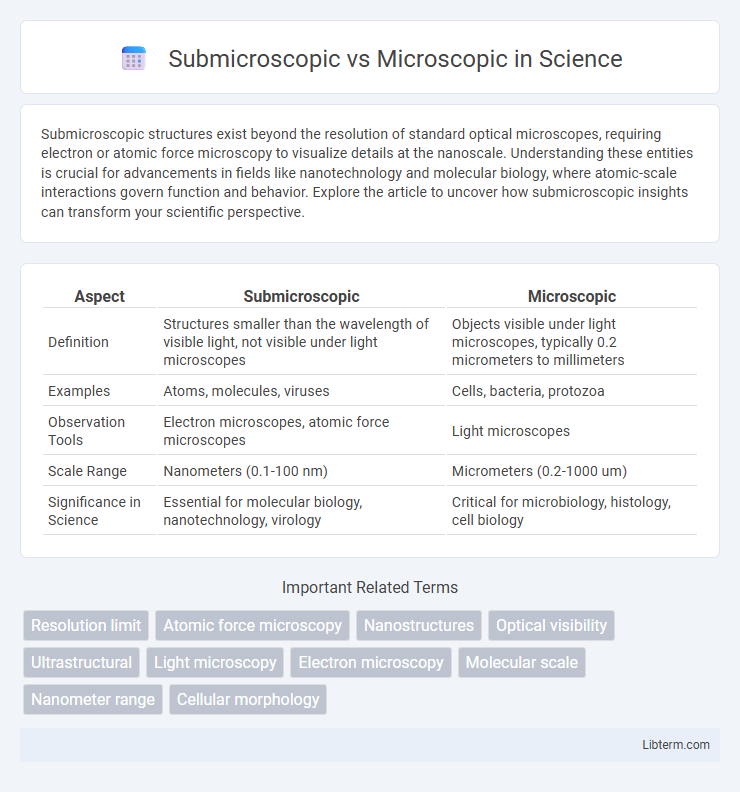
Submicroscopic structures exist beyond the resolution of standard optical microscopes, requiring electron or atomic force microscopy to visualize details at the nanoscale. Understanding these entities is crucial for advancements in fields like nanotechnology and molecular biology, where atomic-scale interactions govern function and behavior. Explore the article to uncover how submicroscopic insights can transform your scientific perspective.
Table of Comparison
| Aspect | Submicroscopic | Microscopic |
|---|---|---|
| Definition | Structures smaller than the wavelength of visible light, not visible under light microscopes | Objects visible under light microscopes, typically 0.2 micrometers to millimeters |
| Examples | Atoms, molecules, viruses | Cells, bacteria, protozoa |
| Observation Tools | Electron microscopes, atomic force microscopes | Light microscopes |
| Scale Range | Nanometers (0.1-100 nm) | Micrometers (0.2-1000 um) |
| Significance in Science | Essential for molecular biology, nanotechnology, virology | Critical for microbiology, histology, cell biology |
Introduction to Submicroscopic and Microscopic Scales
Submicroscopic scale refers to particles and structures smaller than the wavelength of visible light, including atoms, molecules, and electrons, which can only be observed using advanced instruments like electron microscopes or inferred through indirect methods. Microscopic scale encompasses objects visible under optical microscopes, such as cells, bacteria, and tissue structures, ranging from about 1 micrometer to several hundred micrometers. Understanding the distinction between submicroscopic and microscopic scales is essential for fields like chemistry, biology, and materials science, where exploring features at different scales reveals deeper insights into structure and function.
Defining Submicroscopic Structures
Submicroscopic structures refer to components at the molecular or atomic scale, often observed using advanced techniques like electron microscopy or spectroscopy, unlike microscopic structures visible under standard light microscopes. These submicroscopic entities include proteins, DNA, and cellular organelles critical to biochemical processes and cellular functions. Understanding submicroscopic structures enables insights into molecular interactions, mechanisms of disease, and the development of nanotechnology.
What Is Considered Microscopic?
Microscopic refers to objects or structures that are visible only with the aid of a microscope, typically ranging from 0.1 micrometers to several hundred micrometers in size. Examples of microscopic entities include bacteria, single-celled organisms, and cellular components such as organelles. These objects are larger than submicroscopic particles, which are smaller than the wavelength of visible light and require electron microscopy or other advanced techniques for visualization.
Key Differences Between Submicroscopic and Microscopic
Submicroscopic refers to entities smaller than the wavelength of visible light, requiring electron microscopy for visualization, while microscopic involves objects visible under optical microscopes. Submicroscopic structures include atoms and molecules, whereas microscopic structures encompass cells and microorganisms. The primary difference lies in the scale and imaging techniques used, with submicroscopic focusing on nanometer to atomic levels and microscopic on micrometer levels.
Importance in Scientific Research
Submicroscopic analysis reveals atomic and molecular structures invisible to conventional microscopic techniques, enabling breakthroughs in nanotechnology, materials science, and molecular biology. Microscopic methods remain essential for studying cellular and tissue-level phenomena, providing critical insights into physiological processes and disease pathology. Combining both approaches enhances scientific research by bridging scales from molecules to complex biological systems, driving innovation and precision in diagnostics and therapeutic development.
Application in Chemistry and Biology
Submicroscopic analysis reveals atomic and molecular interactions crucial for understanding chemical bonding, reaction mechanisms, and molecular structures in chemistry. Microscopic techniques enable the visualization of cellular components, microorganisms, and tissue structures, facilitating research in biology, such as studying cell morphology and diagnosing diseases. Combining both approaches enhances the analysis of biological macromolecules and the development of pharmaceuticals through detailed structural and functional insights.
Methods of Observation and Analysis
Submicroscopic observation relies on advanced techniques such as electron microscopy, scanning tunneling microscopy (STM), and atomic force microscopy (AFM) to visualize structures at the atomic or molecular scale, enabling analysis beyond the diffraction limit of light. Microscopic methods primarily include optical microscopy, using visible light and lenses to magnify specimens up to 1000 times, suitable for observing cells, bacteria, and larger organelles. Analytical approaches in submicroscopic methods often incorporate spectroscopy and diffraction patterns for detailed material properties, while microscopic techniques focus on staining, phase contrast, and fluorescence for biological specimen visualization.
Challenges in Studying Submicroscopic and Microscopic Matter
Studying submicroscopic matter presents significant challenges due to its scale below the wavelength of visible light, requiring advanced techniques like electron microscopy and atomic force microscopy to visualize and analyze atomic and molecular structures. In contrast, microscopic matter, visible through optical microscopes, allows easier observation but often lacks the resolution needed to explore finer structural details. Both fields face difficulties in sample preparation, imaging resolution, and interpreting complex interactions at different scales, impacting research in materials science, biology, and nanotechnology.
Real-World Examples of Each Scale
Submicroscopic particles include atoms, molecules, and ions, which are essential in chemistry and molecular biology, as seen in DNA strands and protein structures. Microscopic entities like bacteria, cells, and fungi are visible under light microscopes and play crucial roles in medicine, environmental science, and microbiology. Real-world examples of microscopic scale include red blood cells and pollen grains, while submicroscopic scale examples encompass hydrogen atoms and water molecules.
Future Trends and Technological Advances
Future trends in submicroscopic and microscopic imaging focus on enhancing resolution and real-time analysis through advancements in quantum microscopy and electron beam technologies. Integration of AI-powered image processing and nanofabrication techniques promises breakthroughs in materials science, medical diagnostics, and semiconductor manufacturing. Emerging technologies like cryo-electron microscopy and super-resolution fluorescence microscopy continue to push the limits of observing atomic-scale structures and biological processes with unprecedented clarity.
Submicroscopic Infographic

 libterm.com