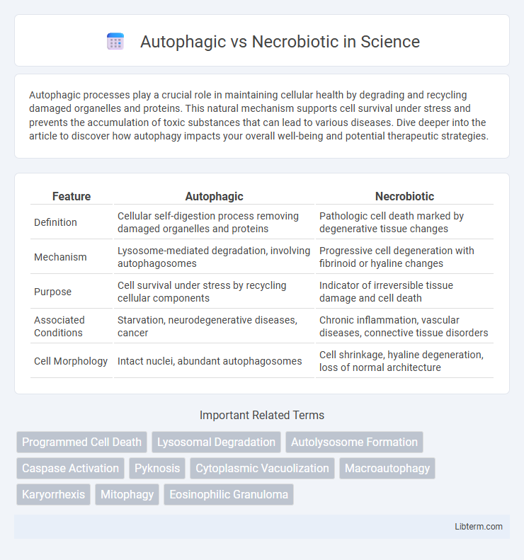
Autophagic processes play a crucial role in maintaining cellular health by degrading and recycling damaged organelles and proteins. This natural mechanism supports cell survival under stress and prevents the accumulation of toxic substances that can lead to various diseases. Dive deeper into the article to discover how autophagy impacts your overall well-being and potential therapeutic strategies.
Table of Comparison
| Feature | Autophagic | Necrobiotic |
|---|---|---|
| Definition | Cellular self-digestion process removing damaged organelles and proteins | Pathologic cell death marked by degenerative tissue changes |
| Mechanism | Lysosome-mediated degradation, involving autophagosomes | Progressive cell degeneration with fibrinoid or hyaline changes |
| Purpose | Cell survival under stress by recycling cellular components | Indicator of irreversible tissue damage and cell death |
| Associated Conditions | Starvation, neurodegenerative diseases, cancer | Chronic inflammation, vascular diseases, connective tissue disorders |
| Cell Morphology | Intact nuclei, abundant autophagosomes | Cell shrinkage, hyaline degeneration, loss of normal architecture |
Introduction to Autophagic and Necrobiotic Processes
Autophagic processes involve the selective degradation of cellular components through lysosomal pathways, essential for maintaining cellular homeostasis and responding to stress by recycling damaged organelles and proteins. Necrobiotic processes, contrastingly, refer to the pathological degeneration and death of cells characterized by structural breakdown and inflammatory responses, often linked to chronic tissue damage. Understanding the distinct mechanisms of autophagy and necrobiosis provides critical insights into cellular survival strategies and pathological cell death.
Defining Autophagic Cell Death
Autophagic cell death is characterized by the extensive degradation of cellular components through the lysosomal machinery, distinct from necrobiotic cell death which involves programmed cellular destruction with inflammation and cellular swelling. Autophagy, a regulated catabolic process, maintains cellular homeostasis by recycling damaged organelles and proteins, and when dysregulated, it leads to autophagic cell death marked by increased autophagosome formation. This form of cell death is vital in various physiological and pathological contexts, including cancer, neurodegeneration, and infection response.
Understanding Necrobiotic Cell Death
Necrobiotic cell death, distinct from autophagic cell death, involves irreversible cellular degeneration characterized by cytoplasmic swelling, nuclear pyknosis, and loss of membrane integrity. Unlike autophagy, which is a regulated process promoting cell survival through degradation of damaged organelles, necrobiosis leads to inflammation due to the release of intracellular contents. Understanding necrobiotic mechanisms is crucial for identifying pathological conditions such as chronic inflammation and degenerative diseases where necrobiotic death predominates.
Molecular Mechanisms of Autophagy
Autophagy is a regulated cellular process involving the degradation of damaged organelles and misfolded proteins through lysosomal pathways, characterized by the formation of autophagosomes. Key molecular mechanisms include the involvement of ATG genes, the ULK1 complex initiating autophagosome formation, and the PI3K complex generating phosphatidylinositol 3-phosphate (PI3P) for membrane nucleation. In contrast, necrobiotic cell death lacks this regulated degradation, occurring via uncontrolled cellular damage and metabolic failure without lysosomal involvement.
Cellular Pathways in Necrobiosis
Necrobiosis involves the programmed degradation of cells through pathways distinct from autophagy, primarily characterized by cell membrane rupture and inflammation. Cellular pathways in necrobiosis include necroptosis, which is regulated by receptor-interacting protein kinases RIPK1 and RIPK3, leading to MLKL-mediated plasma membrane disruption. This process differs from autophagic pathways where lysosomal digestion preserves membrane integrity and promotes cell survival under stress conditions.
Key Differences: Autophagic vs Necrobiotic
Autophagic cell death involves the lysosomal degradation of cellular components through autophagosomes, maintaining membrane integrity and avoiding inflammation, while necrobiotic cell death is characterized by the disintegration of cell structures leading to membrane rupture and triggering inflammation. Autophagy primarily serves as a survival mechanism under stress by recycling damaged organelles, whereas necrobiosis is associated with pathological tissue damage and immune response activation. Key molecular markers differentiate the two processes, with LC3-II and Beclin-1 indicating autophagy, and increased release of damage-associated molecular patterns (DAMPs) signaling necrobiotic cell death.
Biological Significance in Disease and Health
Autophagic cell death plays a crucial role in maintaining cellular homeostasis by degrading damaged organelles and proteins, thereby preventing neurodegenerative diseases and cancer. Necrobiotic processes, characterized by cell death with inflammation, contribute to tissue damage and chronic diseases such as atherosclerosis and rheumatoid arthritis. Understanding the balance between autophagy and necrobiosis is essential for developing targeted therapies that modulate cell death pathways in health and disease.
Diagnostic Markers and Detection Methods
Autophagic cell death is characterized by elevated levels of LC3-II and accumulation of autophagosomes, detectable via immunoblotting and fluorescence microscopy using GFP-LC3 markers. Necrobiotic processes show increased expression of necroptosis markers such as RIPK3 and MLKL, identified through immunohistochemistry and western blot analysis. Distinguishing between autophagic and necrobiotic cell death relies on combining molecular markers with techniques like flow cytometry and electron microscopy to assess ultrastructural changes and specific protein activation.
Therapeutic Implications and Interventions
Autophagic cell death involves the regulated degradation of cellular components, playing a crucial role in maintaining homeostasis and offering therapeutic targets in cancer and neurodegenerative diseases by modulating autophagy pathways with agents like rapamycin and chloroquine. Necrobiotic cell death, characterized by uncontrolled cellular necrosis and inflammation, is often associated with acute tissue injury and chronic inflammatory conditions, necessitating interventions that limit necrosis through antioxidants and anti-inflammatory drugs. Understanding the distinct molecular mechanisms of autophagic versus necrobiotic death aids in developing precise therapies that either promote cellular clearance or prevent excessive tissue damage.
Future Directions in Cell Death Research
Future directions in cell death research emphasize distinguishing autophagic cell death, characterized by cytoplasmic degradation and lysosome involvement, from necrobiotic death marked by irreversible cell membrane rupture and inflammation. Advances in single-cell omics and live-cell imaging technologies enable precise mapping of molecular pathways governing these processes, facilitating targeted therapeutic development. Emerging strategies focus on modulating autophagy for disease treatment while preventing necrobiotic inflammation to improve outcomes in degenerative and inflammatory disorders.
Autophagic Infographic

 libterm.com