Gynecomastia, characterized by the benign enlargement of male breast tissue, can sometimes be mistaken for chest wall tumors due to overlapping physical signs. Accurate diagnosis is essential in distinguishing between these conditions to ensure proper treatment and avoid unnecessary interventions. Explore the article to understand the differences, diagnostic methods, and management options for gynecomastia and chest wall tumors.
Table of Comparison
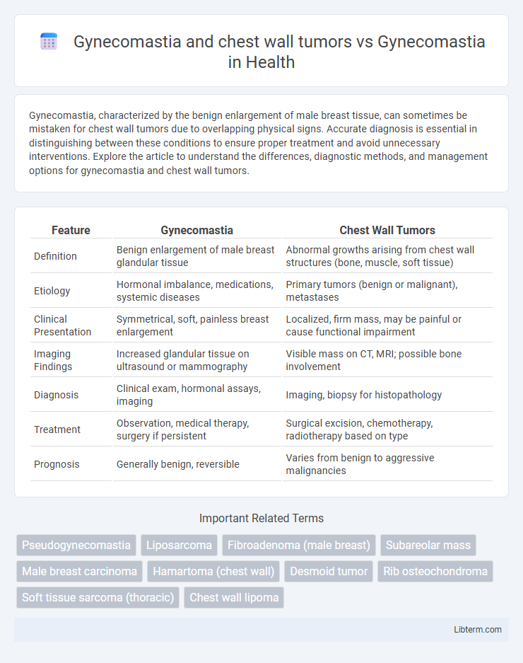
| Feature | Gynecomastia | Chest Wall Tumors |
|---|---|---|
| Definition | Benign enlargement of male breast glandular tissue | Abnormal growths arising from chest wall structures (bone, muscle, soft tissue) |
| Etiology | Hormonal imbalance, medications, systemic diseases | Primary tumors (benign or malignant), metastases |
| Clinical Presentation | Symmetrical, soft, painless breast enlargement | Localized, firm mass, may be painful or cause functional impairment |
| Imaging Findings | Increased glandular tissue on ultrasound or mammography | Visible mass on CT, MRI; possible bone involvement |
| Diagnosis | Clinical exam, hormonal assays, imaging | Imaging, biopsy for histopathology |
| Treatment | Observation, medical therapy, surgery if persistent | Surgical excision, chemotherapy, radiotherapy based on type |
| Prognosis | Generally benign, reversible | Varies from benign to aggressive malignancies |
Understanding Gynecomastia: Definition and Causes
Gynecomastia is the benign enlargement of male breast glandular tissue primarily caused by hormonal imbalances, including increased estrogen or decreased androgen levels. Chest wall tumors, by contrast, are abnormal growths arising from the bones, muscles, or soft tissues of the chest and can mimic gynecomastia but require distinct diagnostic evaluation to differentiate. Understanding gynecomastia involves recognizing its etiologies such as puberty, medication side effects, systemic diseases, and distinguishing it from pathological masses like chest wall tumors through clinical examination and imaging studies.
What Are Chest Wall Tumors? An Overview
Chest wall tumors are abnormal growths that arise from the bones, muscles, or connective tissues of the chest wall and can be benign or malignant. Unlike gynecomastia, which involves the benign enlargement of male breast glandular tissue, chest wall tumors may present with a palpable mass, pain, or deformity and often require imaging and biopsy for diagnosis. Accurate differentiation between gynecomastia and chest wall tumors is essential due to their distinct pathological origins and treatment strategies.
Key Differences Between Gynecomastia and Chest Wall Tumors
Gynecomastia is characterized by benign enlargement of male breast glandular tissue, whereas chest wall tumors represent abnormal growths originating from muscles, bones, or other chest components and may be benign or malignant. Key differences include the presence of firm, often tender breast tissue in gynecomastia compared to potentially palpable masses with variable consistency in chest wall tumors. Imaging techniques such as mammography and ultrasound help differentiate gynecomastia's diffuse glandular proliferation from the localized, irregular masses typical of chest wall tumors.
Clinical Presentation: Gynecomastia vs. Chest Wall Tumors
Gynecomastia typically presents as a symmetric, painless, rubbery enlargement of the male breast tissue, often bilateral and centered around the areola without associated skin changes. In contrast, chest wall tumors usually manifest as a localized mass, which may be firm or hard, potentially accompanied by pain, tenderness, or skin ulceration, and are often unilateral. Clinical examination differentiates gynecomastia by its diffuse glandular proliferation, whereas chest wall tumors involve deeper structures such as ribs or muscles with possible systemic symptoms like weight loss or fatigue.
Diagnostic Approaches for Gynecomastia and Chest Wall Masses
Diagnostic approaches for gynecomastia primarily involve clinical examination, hormonal evaluation including serum testosterone, estradiol, and prolactin levels, and imaging studies such as ultrasound to assess glandular proliferation. In contrast, chest wall tumors require advanced imaging modalities like MRI or CT scans to evaluate lesion size, extent, and involvement of adjacent structures, alongside biopsy for histopathological diagnosis. Differentiating gynecomastia from chest wall masses relies heavily on combining physical findings with targeted imaging and laboratory tests to ensure accurate diagnosis and appropriate management.
Imaging Techniques: Ultrasound, Mammography, and MRI
Imaging techniques such as ultrasound, mammography, and MRI play critical roles in differentiating gynecomastia from chest wall tumors. Ultrasound provides high-resolution images to distinguish glandular proliferation in gynecomastia from solid or cystic masses typical of tumors. Mammography assists in identifying asymmetric breast tissue and calcifications, while MRI offers detailed soft tissue contrast to evaluate tumor extent and vascularity, enhancing diagnostic accuracy.
Treatment Options for Gynecomastia
Treatment options for gynecomastia primarily include medical therapies such as selective estrogen receptor modulators (SERMs) like tamoxifen and aromatase inhibitors, while chest wall tumors often require surgical excision followed by oncologic evaluation. In gynecomastia cases resistant to medication or those with persistent breast tissue causing psychological distress, liposuction or subcutaneous mastectomy are effective surgical treatments. Differentiating gynecomastia from chest wall tumors through imaging and biopsy ensures targeted treatment, as tumors may necessitate chemotherapy or radiotherapy alongside surgery.
Management Strategies for Chest Wall Tumors
Management strategies for chest wall tumors differ significantly from those for gynecomastia due to the malignant potential and structural involvement of tumors. Surgical resection combined with reconstructive techniques is the primary approach for chest wall tumors, often supplemented by chemotherapy or radiotherapy depending on tumor histology and staging. In contrast, gynecomastia typically requires conservative management, hormonal therapy, or reduction mammoplasty when symptomatic or persistent.
Risks and Complications: Differentiating Malignancy
Gynecomastia primarily involves benign proliferation of male breast glandular tissue, whereas chest wall tumors may represent malignant growths with potential for local invasion and metastasis. Differentiating malignancy is critical, as chest wall tumors pose higher risks including pain, ulceration, and systemic complications, necessitating prompt diagnostic imaging and biopsy. Misdiagnosis of a malignant chest wall tumor as gynecomastia can delay treatment and worsen prognosis, emphasizing thorough clinical examination and histopathological evaluation.
Prognosis and Follow-Up: Ensuring Optimal Patient Outcomes
Gynecomastia, characterized by benign glandular breast tissue enlargement in males, generally has an excellent prognosis with minimal risk of malignancy, requiring routine clinical follow-up to monitor for changes. In contrast, chest wall tumors, which can be benign or malignant, demand more intensive evaluation and tailored follow-up protocols involving imaging studies and possible biopsy to ensure early detection of recurrence or progression. Effective differentiation and vigilant monitoring are critical for optimizing patient outcomes, guiding timely interventions, and improving overall prognosis.
Gynecomastia and chest wall tumors Infographic