Enophthalmos is the posterior displacement of the eyeball within the orbit, often caused by trauma, orbital fractures, or conditions leading to volume loss in the orbit. This condition can result in noticeable asymmetry, vision disturbances, and cosmetic concerns that may affect your quality of life. Explore the following article to understand the causes, symptoms, and treatment options for enophthalmos.
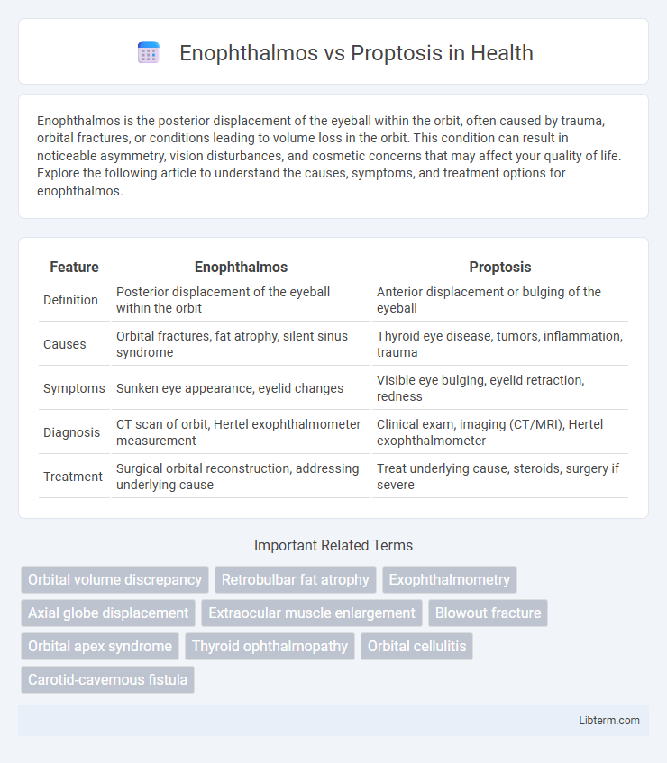
Table of Comparison
| Feature | Enophthalmos | Proptosis |
|---|---|---|
| Definition | Posterior displacement of the eyeball within the orbit | Anterior displacement or bulging of the eyeball |
| Causes | Orbital fractures, fat atrophy, silent sinus syndrome | Thyroid eye disease, tumors, inflammation, trauma |
| Symptoms | Sunken eye appearance, eyelid changes | Visible eye bulging, eyelid retraction, redness |
| Diagnosis | CT scan of orbit, Hertel exophthalmometer measurement | Clinical exam, imaging (CT/MRI), Hertel exophthalmometer |
| Treatment | Surgical orbital reconstruction, addressing underlying cause | Treat underlying cause, steroids, surgery if severe |
Introduction to Enophthalmos and Proptosis
Enophthalmos is characterized by the posterior displacement of the eyeball within the orbit, often caused by trauma, orbital fat atrophy, or silent sinus syndrome. Proptosis, also known as exophthalmos, refers to the forward bulging of the eye, commonly associated with thyroid eye disease, orbital tumors, or inflammation. Both conditions require careful clinical evaluation and imaging studies to determine underlying etiology and guide appropriate treatment.
Defining Enophthalmos: Key Characteristics
Enophthalmos is characterized by the posterior displacement of the eyeball within the orbit, often caused by trauma, orbital fractures, or conditions leading to orbital fat atrophy. It contrasts with proptosis, which involves the forward protrusion of the eye due to increased orbital volume or mass effect. Key features of enophthalmos include sunken appearance of the eye, relative enophthalmos compared to the contralateral side, and possible associated orbital rim deformities visible on CT imaging.
Understanding Proptosis: Main Features
Proptosis refers to the abnormal forward displacement of the eyeball, often caused by conditions such as thyroid eye disease, orbital tumors, or inflammation. This protrusion can lead to symptoms including eye dryness, irritation, and impaired eyelid closure, making it distinct from enophthalmos, which involves the recession of the eyeball into the orbit. Accurate diagnosis of proptosis relies on clinical evaluation and imaging techniques like CT or MRI scans to assess the underlying orbital pathology.
Anatomical Differences Between Enophthalmos and Proptosis
Enophthalmos is characterized by the posterior displacement of the eyeball within the orbit, often caused by a reduction in orbital volume due to trauma, fat atrophy, or structural abnormalities. Proptosis involves the anterior protrusion of the globe beyond the orbital rim, frequently resulting from increased orbital content such as tumors, inflammation, or vascular malformations. Anatomical differences are centered on the orbital volume and contents, with enophthalmos linked to decreased orbital space and proptosis associated with expanded orbital tissue or mass effect.
Common Causes of Enophthalmos
Enophthalmos is characterized by the posterior displacement of the eyeball within the orbit, often caused by orbital trauma, silent sinus syndrome, or orbital fat atrophy. Unlike proptosis, which involves the anterior protrusion of the eye often due to thyroid eye disease or orbital tumors, enophthalmos commonly arises from orbital fractures leading to increased orbital volume or scarring. Recognizing these underlying conditions is crucial for accurate diagnosis and effective management of enophthalmos.
Typical Etiologies of Proptosis
Proptosis, also known as exophthalmos, is typically caused by thyroid eye disease (Graves' orbitopathy), orbital cellulitis, tumors such as orbital lymphoma or meningioma, and vascular conditions like carotid-cavernous fistula or orbital varices. In contrast, enophthalmos often results from trauma causing orbital floor fractures or silent sinus syndrome leading to posterior displacement of the globe. Understanding these etiologies is crucial for accurate diagnosis and targeted treatment of orbital abnormalities.
Clinical Presentation and Symptoms
Enophthalmos is characterized by posterior displacement of the eyeball within the orbit, often presenting with a sunken appearance, eyelid tightness, and decreased orbital volume frequently caused by trauma or orbital fat loss. Proptosis involves anterior displacement of the eyeball, manifesting as bulging eyes, eyelid retraction, and potential exposure keratopathy, commonly associated with thyroid eye disease or orbital tumors. Clinical symptoms for enophthalmos include diplopia and enophthalmos-related aesthetic concerns, while proptosis often presents with vision changes, pain, and ocular dryness due to increased ocular exposure.
Diagnostic Techniques and Tools
Diagnostic techniques for enophthalmos primarily include clinical examination, Hertel exophthalmometry to measure posterior displacement of the globe, and orbital imaging such as CT or MRI to assess orbital volume and structural anomalies. Proptosis assessment relies on Hertel or Naugle exophthalmometry for quantifying anterior displacement and orbital imaging to identify causes like tumors, inflammation, or vascular anomalies. Ultrasound Doppler and angiography further aid in diagnosing vascular causes of proptosis, while biopsy may be necessary to confirm tumor-related etiologies.
Treatment and Management Approaches
Treatment and management of enophthalmos primarily target the underlying cause, such as orbital fractures, trauma, or chronic sinus disease, often requiring surgical intervention like orbital floor reconstruction to restore orbital volume and position. In contrast, proptosis management focuses on addressing the etiology, ranging from corticosteroids or radiotherapy for thyroid eye disease to surgical decompression or tumor removal in cases of orbital tumors or inflammation. Both conditions necessitate multidisciplinary care involving ophthalmologists, radiologists, and sometimes neurosurgeons to optimize functional and cosmetic outcomes.
Prognosis and Patient Outcomes
Enophthalmos, characterized by the posterior displacement of the eyeball within the orbit, often indicates underlying orbital volume loss or structural damage and can result in persistent cosmetic deformity and impaired ocular function if untreated. Proptosis, the anterior protrusion of the eye, typically signals active orbital inflammation, tumors, or Graves' disease, and prognosis depends heavily on timely diagnosis and management of the underlying cause to prevent vision loss or optic neuropathy. Patient outcomes vary: enophthalmos prognosis worsens with delayed intervention leading to permanent visual and aesthetic issues, while proptosis outcomes improve significantly with early targeted therapy reducing the risk of complications such as exposure keratopathy or compressive optic neuropathy.
Enophthalmos Infographic