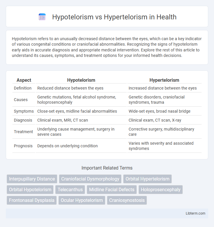
Hypotelorism refers to an unusually decreased distance between the eyes, which can be a key indicator of various congenital conditions or craniofacial abnormalities. Recognizing the signs of hypotelorism early aids in accurate diagnosis and appropriate medical intervention. Explore the rest of this article to understand its causes, symptoms, and treatment options for your informed health decisions.
Table of Comparison
| Aspect | Hypotelorism | Hypertelorism |
|---|---|---|
| Definition | Reduced distance between the eyes | Increased distance between the eyes |
| Causes | Genetic mutations, fetal alcohol syndrome, holoprosencephaly | Genetic disorders, craniofacial syndromes, trauma |
| Symptoms | Close-set eyes, midline facial abnormalities | Wide-set eyes, broad nasal bridge |
| Diagnosis | Clinical exam, MRI, CT scan | Clinical exam, CT scan, X-ray |
| Treatment | Underlying cause management, surgery in severe cases | Corrective surgery, multidisciplinary care |
| Prognosis | Depends on underlying condition | Varies with severity and associated syndromes |
Understanding Hypotelorism and Hypertelorism
Hypotelorism refers to an abnormally decreased distance between the eyes, often associated with developmental anomalies such as holoprosencephaly or craniofacial syndromes. Hypertelorism describes an increased distance between the orbits, commonly linked to conditions like craniofacial dysplasia, including Crouzon or Apert syndrome. Accurate measurement of the interorbital distance using imaging techniques like CT or MRI is essential for diagnosing and differentiating these craniofacial abnormalities.
Key Anatomical Differences
Hypotelorism is characterized by an abnormally decreased distance between the orbits, resulting in closely spaced eyes, whereas hypertelorism involves an increased interorbital distance causing widely spaced eyes. Key anatomical differences include the positioning of the medial orbital walls, with hypotelorism showing closer and sometimes fused medial canthi, while hypertelorism features lateral displacement of the orbits. Craniofacial abnormalities affecting the ethmoid bone and nasal bridge morphology are often associated, with hypotelorism usually linked to midline facial hypoplasia and hypertelorism related to frontonasal dysplasia.
Embryological Development and Causes
Hypotelorism and hypertelorism result from disrupted embryological development of the craniofacial region, specifically involving abnormal migration or growth of neural crest cells affecting the formation of the orbits. Hypotelorism, characterized by abnormally decreased interorbital distance, typically arises from early midline defects such as holoprosencephaly or impaired optic vesicle separation. Hypertelorism, marked by increased interorbital distance, is commonly caused by frontonasal dysplasia, craniosynostosis syndromes, or excessive growth of the ethmoid bone during facial development.
Clinical Presentation and Diagnosis
Hypotelorism is characterized by an abnormally decreased distance between the orbits, often associated with midline facial anomalies and brain malformations, while hypertelorism presents as an increased interorbital distance frequently linked to craniofacial syndromes and developmental abnormalities. Clinical diagnosis involves precise measurement of interorbital distances using imaging modalities such as MRI or CT scans, supplemented by craniofacial anthropometric assessments to determine deviations from normative values. Genetic testing and detailed neurological evaluations support differential diagnosis and guide management strategies for associated syndromic conditions.
Associated Syndromes and Conditions
Hypotelorism, characterized by abnormally decreased distance between the eyes, is commonly associated with syndromes such as Holoprosencephaly and Trisomy 13 (Patau syndrome). In contrast, hypertelorism, defined by an increased distance between the orbits, is frequently observed in conditions like Apert syndrome, Crouzon syndrome, and Waardenburg syndrome. Accurate diagnosis of these orbital spacing abnormalities helps guide genetic testing and management for underlying craniofacial and developmental disorders.
Diagnostic Imaging and Measurement Techniques
Hypotelorism and hypertelorism are congenital conditions characterized by decreased and increased interorbital distances, respectively, diagnosed primarily through advanced imaging techniques such as computed tomography (CT) and magnetic resonance imaging (MRI). Precise measurement of the interorbital distance involves assessing the distance between the medial orbital walls (for hypotelorism) or the orbital rims (for hypertelorism) on axial CT scans, with normative data stratified by age and sex serving as a comparative standard. Three-dimensional reconstruction and cephalometric analysis enhance diagnostic accuracy, aiding in surgical planning and differentiation from other craniofacial anomalies.
Treatment Options and Management Strategies
Hypotelorism, characterized by abnormally decreased distance between the eyes, often requires multidisciplinary treatment including surgical interventions such as orbital medialization to correct ocular spacing and improve function and appearance. Hypertelorism involves an increased interorbital distance typically managed through craniofacial surgery techniques like box osteotomy or facial bipartition to reposition the orbits and restore facial symmetry. Both conditions benefit from early diagnosis, individualized surgical planning, and postoperative monitoring to optimize functional and aesthetic outcomes.
Prognosis and Long-Term Outcomes
Hypotelorism, characterized by abnormally decreased distance between the eyes, often indicates underlying midline brain anomalies, which may result in developmental delays and complex neurological issues, impacting long-term cognitive and motor outcomes. Hypertelorism, defined by increased interorbital distance, is frequently associated with craniofacial syndromes like craniofrontonasal dysplasia and may require surgical intervention to improve functional and aesthetic prognosis, with variable effects on psychosocial development. Long-term outcomes for both conditions depend on the severity of associated anomalies and the timeliness of multidisciplinary management, including neurology, genetics, and reconstructive surgery.
Pediatric vs. Adult Considerations
Hypotelorism, characterized by abnormally decreased distance between the orbits, poses significant diagnostic challenges in pediatric cases due to ongoing craniofacial development, whereas hypertelorism involves an increased interorbital distance that often correlates with complex syndromic conditions more commonly identified in adults. Pediatric management focuses on early detection through imaging modalities like MRI and 3D CT scans to guide potential surgical interventions during critical growth periods, while adult assessment emphasizes functional and aesthetic outcomes with tailored reconstructive procedures. Understanding age-specific anatomical and developmental variations is crucial for effective treatment planning and improving long-term prognosis in patients presenting with these orbital distance anomalies.
Current Research and Future Directions
Current research on hypotelorism and hypertelorism emphasizes genetic and embryological pathways influencing craniofacial development, with studies utilizing advanced imaging techniques such as 3D CT scans and MRI for precise phenotypic characterization. Emerging molecular analyses target gene mutations like SHH, FGFR, and GLI3, aiming to unravel the distinct mechanisms driving orbital distance anomalies. Future directions include gene therapy approaches, personalized medicine, and improved diagnostic protocols integrating AI algorithms to enhance early detection and tailored treatment strategies for patients with orbital spacing disorders.
Hypotelorism Infographic

 libterm.com