Orbital hypertelorism is a rare craniofacial anomaly characterized by an abnormally increased distance between the orbits, often linked to genetic syndromes or developmental disturbances. This condition can affect your appearance and may require surgical intervention to improve both function and aesthetics. Explore the following article to learn about causes, diagnosis, and treatment options for orbital hypertelorism.
Table of Comparison
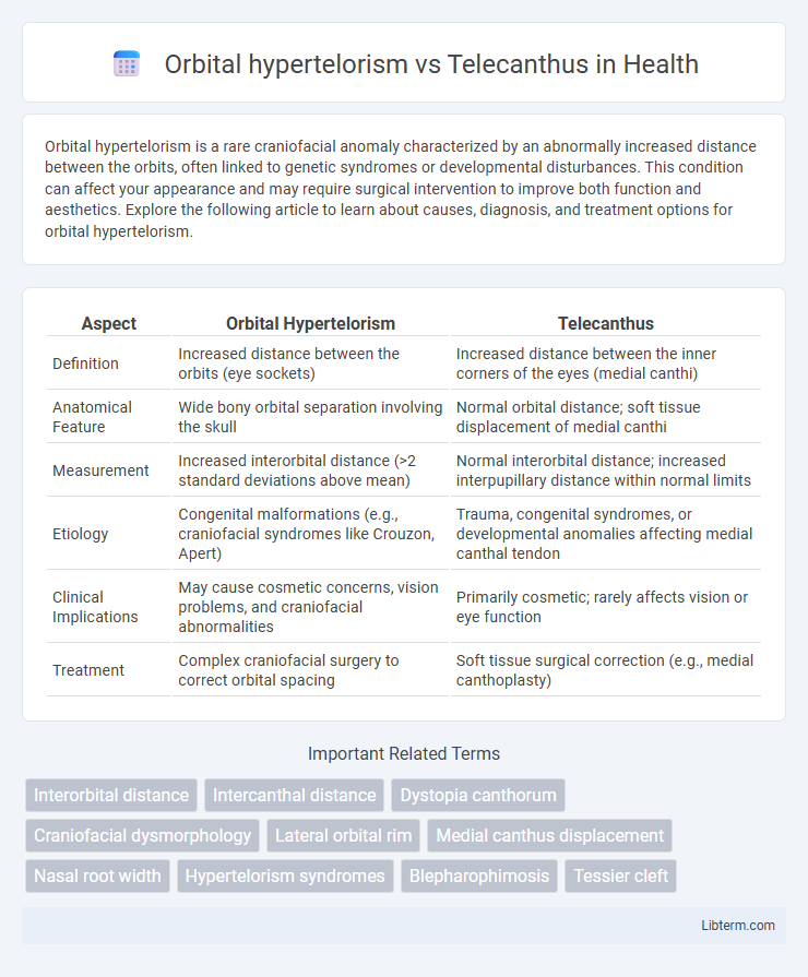
| Aspect | Orbital Hypertelorism | Telecanthus |
|---|---|---|
| Definition | Increased distance between the orbits (eye sockets) | Increased distance between the inner corners of the eyes (medial canthi) |
| Anatomical Feature | Wide bony orbital separation involving the skull | Normal orbital distance; soft tissue displacement of medial canthi |
| Measurement | Increased interorbital distance (>2 standard deviations above mean) | Normal interorbital distance; increased interpupillary distance within normal limits |
| Etiology | Congenital malformations (e.g., craniofacial syndromes like Crouzon, Apert) | Trauma, congenital syndromes, or developmental anomalies affecting medial canthal tendon |
| Clinical Implications | May cause cosmetic concerns, vision problems, and craniofacial abnormalities | Primarily cosmetic; rarely affects vision or eye function |
| Treatment | Complex craniofacial surgery to correct orbital spacing | Soft tissue surgical correction (e.g., medial canthoplasty) |
Introduction to Orbital Hypertelorism and Telecanthus
Orbital hypertelorism is a craniofacial condition characterized by an abnormally increased distance between the bony orbits, measured as an interorbital distance exceeding normative values for age and sex. Telecanthus refers to an increased distance between the medial canthi of the eyelids, with a normal bony orbital structure and interorbital distance. Differentiating these conditions is crucial for accurate diagnosis and surgical planning since orbital hypertelorism involves skeletal abnormalities, whereas telecanthus is a soft tissue displacement without true orbital widening.
Defining Orbital Hypertelorism
Orbital hypertelorism is a congenital condition characterized by an abnormally increased distance between the orbits, specifically the bony sockets of the eyes, resulting in widely spaced eyes. This differs from telecanthus, which involves an increased distance between the medial canthi (inner corners) of the eyelids without a corresponding increase in the bony orbital distance. Orbital hypertelorism often indicates underlying craniofacial anomalies and requires precise measurement of the interpupillary distance and interorbital distance for accurate diagnosis.
What is Telecanthus?
Telecanthus is a condition characterized by an increased distance between the medial canthi of the eyes, where the inner corners of the eyelids are abnormally spaced apart while the interpupillary distance remains normal. Unlike orbital hypertelorism, which involves an increased distance between the bony orbits, telecanthus primarily affects the soft tissue positioning of the eyelids without altering the underlying orbital bones. This distinction is crucial for accurate diagnosis and appropriate surgical planning in craniofacial abnormalities.
Key Clinical Differences
Orbital hypertelorism involves an increased distance between the bony orbits, typically measured by a widened interorbital distance exceeding normal age-related values, whereas telecanthus refers to an increased distance between the medial canthi with a normal bony orbital distance. Key clinical differences include orbital hypertelorism presenting with both skeletal and soft tissue changes, often visible on craniofacial imaging, while telecanthus affects only the soft tissue positioning without underlying bone displacement. Telecanthus commonly occurs in conditions like Waardenburg syndrome, whereas orbital hypertelorism is associated with craniofacial syndromes such as frontonasal dysplasia.
Etiology and Associated Syndromes
Orbital hypertelorism is characterized by an increased distance between the orbits due to bony abnormalities, often linked to craniofacial syndromes such as Crouzon, Apert, and Waardenburg syndromes. Telecanthus involves a widened distance between the medial canthi with normal orbital spacing, commonly associated with conditions like Waardenburg syndrome, Down syndrome, and chromosome 22q11.2 deletion syndrome. Both conditions arise from developmental anomalies during embryogenesis, affecting craniofacial bone and soft tissue structures.
Diagnostic Approaches
Orbital hypertelorism is diagnosed through precise craniofacial measurements like increased interpupillary distance and orbital width, often confirmed by 3D CT imaging, which distinguishes the true lateral displacement of the orbits. Telecanthus is characterized by an increased distance between the medial canthi with normal orbital spacing, typically evaluated via direct anthropometric assessment and corroborated by imaging to rule out orbital malposition. Differential diagnosis relies on combining clinical metrics with radiological evidence to distinguish between lateral orbital displacement and medial canthal soft tissue anomalies.
Clinical Assessment and Measurements
Orbital hypertelorism involves an increased distance between the orbits with a widened interorbital bone, assessed using the orbital index and frontozygomatic measurements on CT or MRI scans. Telecanthus is characterized by an increased distance between the medial canthi while the bony orbital distance remains normal, measured clinically by the inner canthal distance compared to age-matched norms. Precise anthropometric measurements, including interpupillary distance and nasal root position, are critical in differentiating between these two conditions during clinical assessment.
Treatment and Management Options
Orbital hypertelorism treatment primarily involves extensive craniofacial surgery aimed at repositioning the orbits to correct the widened distance between the eyes, often performed in early childhood to optimize functional and aesthetic outcomes. Telecanthus management focuses on less invasive surgical procedures, such as medial canthoplasty, to reduce the increased distance between the inner canthi while preserving orbital position. Both conditions require multidisciplinary approaches involving ophthalmologists, plastic surgeons, and sometimes neurosurgeons for optimal functional and cosmetic rehabilitation.
Prognosis and Outcomes
Orbital hypertelorism, characterized by an increased distance between the orbital bones, often requires surgical correction to improve facial symmetry and function, with prognosis depending on the severity and associated anomalies. Telecanthus, marked by increased distance between the medial canthi but normal orbital width, generally presents better outcomes with less complex treatment and minimal impact on vision or development. Both conditions benefit from early diagnosis and multidisciplinary management to optimize aesthetic and functional results.
Summary and Future Perspectives
Orbital hypertelorism is characterized by an increased distance between the bony orbits, whereas telecanthus involves an increased distance between the medial canthi without alteration in the bony orbit spacing. Advances in 3D imaging and genetic analysis improve diagnostic precision, enabling tailored surgical interventions for craniofacial anomalies. Future perspectives emphasize integrating molecular genetics with innovative reconstructive techniques to enhance functional and aesthetic outcomes in affected individuals.
Orbital hypertelorism Infographic