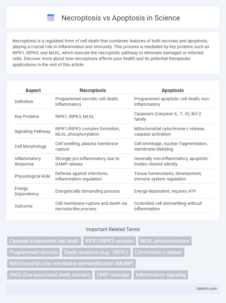
Necroptosis is a regulated form of cell death that combines features of both necrosis and apoptosis, playing a crucial role in inflammation and immunity. This process is mediated by key proteins such as RIPK1, RIPK3, and MLKL, which execute the necroptotic pathway to eliminate damaged or infected cells. Discover more about how necroptosis affects your health and its potential therapeutic applications in the rest of this article.
Table of Comparison
| Aspect | Necroptosis | Apoptosis |
|---|---|---|
| Definition | Programmed necrotic cell death; inflammatory | Programmed apoptotic cell death; non-inflammatory |
| Key Proteins | RIPK1, RIPK3, MLKL | Caspases (Caspase-3, -7, -9), Bcl-2 family |
| Signaling Pathway | RIPK1/RIPK3 complex formation, MLKL phosphorylation | Mitochondrial cytochrome c release, caspase activation |
| Cell Morphology | Cell swelling, plasma membrane rupture | Cell shrinkage, nuclear fragmentation, membrane blebbing |
| Inflammatory Response | Strongly pro-inflammatory due to DAMP release | Generally non-inflammatory, apoptotic bodies cleared silently |
| Physiological Role | Defense against infections, inflammation regulation | Tissue homeostasis, development, immune system regulation |
| Energy Dependency | Energetically demanding process | Energy-dependent, requires ATP |
| Outcome | Cell membrane rupture and death via necrosis-like process | Controlled cell dismantling without inflammation |
Introduction to Programmed Cell Death
Programmed cell death encompasses necroptosis and apoptosis, two distinct yet regulated mechanisms vital for cellular homeostasis. Apoptosis features energy-dependent, caspase-mediated processes leading to cell shrinkage and DNA fragmentation without eliciting inflammation. Necroptosis, regulated by receptor-interacting protein kinases RIPK1 and RIPK3, triggers a controlled necrotic pathway causing cell swelling and membrane rupture, often associated with inflammatory responses.
Defining Necroptosis: Mechanisms and Triggers
Necroptosis is a regulated form of necrotic cell death triggered by specific stress signals such as tumor necrosis factor (TNF), viral infections, or cellular damage, involving the activation of receptor-interacting protein kinases RIPK1 and RIPK3. This programmed necrosis differs from apoptosis by its reliance on the necrosome complex formation, leading to plasma membrane rupture and inflammation rather than the caspase-driven, non-inflammatory cell dismantling seen in apoptosis. Key molecular events in necroptosis include phosphorylation of mixed lineage kinase domain-like protein (MLKL), which translocates to the membrane causing cell lysis.
Apoptosis: Process and Key Features
Apoptosis is a programmed cell death process characterized by energy-dependent biochemical events leading to cell shrinkage, chromatin condensation, and DNA fragmentation. Key features include the activation of caspases, particularly initiator caspases (caspase-8 and caspase-9) and executioner caspases (caspase-3 and caspase-7), which orchestrate cell dismantling without eliciting inflammation. This tightly regulated mechanism maintains tissue homeostasis and eliminates damaged or potentially harmful cells, contrasting with necroptosis, which is a pro-inflammatory, caspase-independent form of regulated necrosis.
Molecular Pathways: Necroptosis vs Apoptosis
Necroptosis and apoptosis are distinct programmed cell death pathways involving different molecular mechanisms; apoptosis primarily activates caspases such as caspase-3 and caspase-8, leading to controlled cell dismantling without inflammation. Necroptosis relies on receptor-interacting protein kinases RIPK1, RIPK3, and the effector protein MLKL, which disrupt plasma membrane integrity and trigger inflammatory responses. Understanding the differential activation of these pathways is critical for therapeutic strategies in diseases including cancer, neurodegeneration, and inflammatory conditions.
Cellular Morphology Differences
Necroptosis is characterized by cellular swelling, plasma membrane rupture, and organelle disintegration, contrasting with apoptosis, which displays cell shrinkage, chromatin condensation, and formation of apoptotic bodies. In necroptosis, loss of membrane integrity leads to an inflammatory response, while apoptosis maintains membrane integrity, preventing inflammation. These distinct morphological features reflect the different molecular pathways and physiological outcomes of necroptosis and apoptosis.
Biological Functions and Physiological Roles
Necroptosis is a programmed form of necrotic cell death that plays a crucial role in immune response regulation and inflammation, often activated when apoptosis is inhibited. Apoptosis is a tightly regulated process essential for maintaining tissue homeostasis, eliminating damaged or dangerous cells without triggering an inflammatory response. Both pathways contribute to development, immune defense, and disease progression, but necroptosis is associated with pathological conditions involving inflammation, while apoptosis primarily supports normal cell turnover and organismal health.
Implications in Disease and Pathology
Necroptosis and apoptosis represent two distinct forms of programmed cell death with critical implications in disease and pathology. Necroptosis, characterized by the activation of RIPK1, RIPK3, and MLKL, plays a pivotal role in inflammatory diseases, neurodegeneration, and cancer resistance by triggering inflammation and immune responses. In contrast, apoptosis involves caspase-mediated cell dismantling, maintaining tissue homeostasis and preventing autoimmune disorders, with dysregulation linked to cancer development and chronic infections.
Therapeutic Targeting: Opportunities and Challenges
Necroptosis and apoptosis represent distinct programmed cell death pathways that serve as promising therapeutic targets in cancer and neurodegenerative diseases. Targeting necroptosis involves modulating key regulators like RIPK1, RIPK3, and MLKL to induce inflammatory cell death in apoptosis-resistant tumors, while apoptosis targeting focuses on caspase activation and Bcl-2 family proteins to selectively trigger cell suicide. Challenges include balancing therapeutic efficacy with potential inflammation from necroptosis activation and overcoming resistance mechanisms in apoptosis, necessitating precise modulation of these pathways for effective clinical applications.
Diagnostic Markers and Detection Methods
Necroptosis is primarily characterized by the activation of receptor-interacting protein kinases RIPK1 and RIPK3, and the mixed lineage kinase domain-like protein (MLKL) phosphorylation serves as a specific diagnostic marker, detected using Western blotting or immunohistochemistry. Apoptosis involves caspase activation, particularly caspase-3 cleavage, and the externalization of phosphatidylserine, with detection methods including TUNEL assay, flow cytometry using Annexin V staining, and caspase activity assays. Differentiating necroptosis from apoptosis relies on combined analysis of molecular markers and morphological assessment under microscopy to identify cell membrane integrity loss in necroptosis versus cell shrinkage and nuclear fragmentation in apoptosis.
Future Directions in Cell Death Research
Future directions in cell death research emphasize dissecting the molecular crosstalk between necroptosis and apoptosis to develop targeted therapies for inflammatory and degenerative diseases. Advanced single-cell sequencing and live-cell imaging technologies are being leveraged to precisely map the spatiotemporal dynamics and regulatory checkpoints of these pathways. Investigating necroptosis triggers and apoptosis resistance mechanisms will facilitate novel drug discovery aimed at modulating pathological cell death in cancer and neurodegeneration.
Necroptosis Infographic

 libterm.com