Pyknosis is the irreversible condensation of chromatin in the nucleus of a cell undergoing apoptosis or necrosis, resulting in a shrunken and intensely stained nucleus. This process is a critical marker for identifying cell death in histopathology and aids in diagnosing various diseases. Explore the rest of the article to understand how pyknosis impacts cellular health and its diagnostic significance in medical research.
Table of Comparison
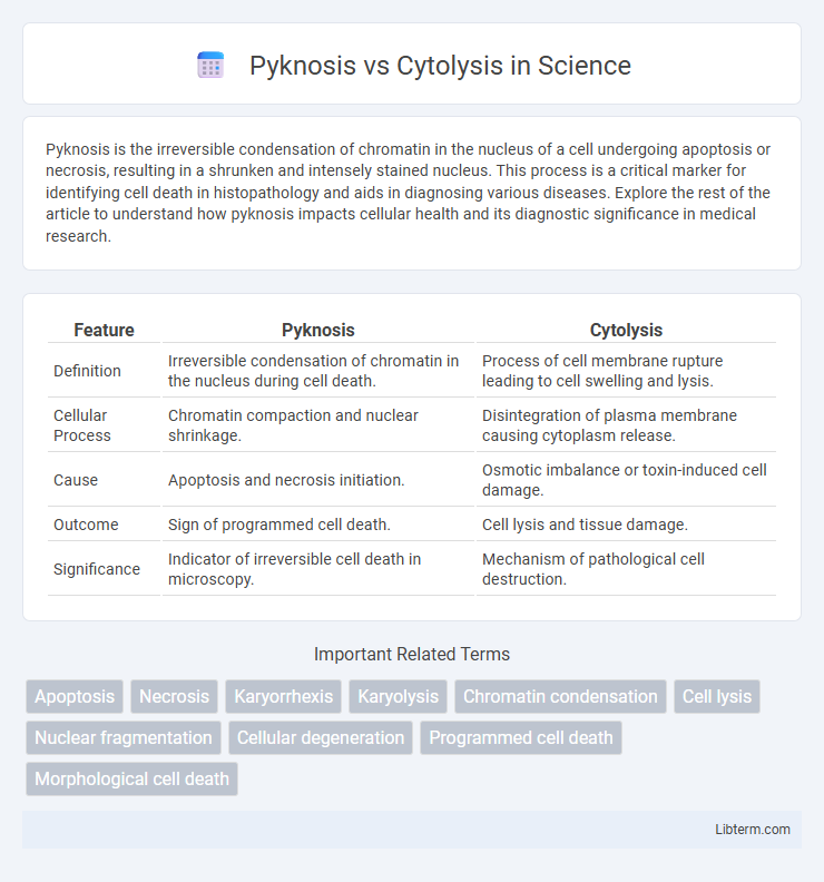
| Feature | Pyknosis | Cytolysis |
|---|---|---|
| Definition | Irreversible condensation of chromatin in the nucleus during cell death. | Process of cell membrane rupture leading to cell swelling and lysis. |
| Cellular Process | Chromatin compaction and nuclear shrinkage. | Disintegration of plasma membrane causing cytoplasm release. |
| Cause | Apoptosis and necrosis initiation. | Osmotic imbalance or toxin-induced cell damage. |
| Outcome | Sign of programmed cell death. | Cell lysis and tissue damage. |
| Significance | Indicator of irreversible cell death in microscopy. | Mechanism of pathological cell destruction. |
Introduction to Pyknosis and Cytolysis
Pyknosis is a form of cellular death characterized by nuclear condensation and shrinkage, indicating irreversible cell damage during apoptosis. Cytolysis involves the breakdown of the cell membrane, leading to the loss of cellular contents and cell bursting, commonly observed in necrotic cell death. Both processes reflect distinct mechanisms of cell demise with pyknosis signaling controlled apoptosis and cytolysis representing uncontrolled necrosis.
Defining Pyknosis: Key Features
Pyknosis is characterized by the irreversible condensation of chromatin in the nucleus, resulting in a shrunken, dense, and intensely basophilic appearance under microscopy. This key feature marks the early stage of apoptosis or necrosis, distinguishing pyknosis from cytolysis, which involves the swelling and rupture of the cell membrane leading to cell lysis. Pyknosis serves as a critical morphological indicator of cell death, contrasting with cytolysis that reflects membrane integrity loss and cytoplasmic disruption.
Understanding Cytolysis: An Overview
Cytolysis is the process where a cell bursts due to excessive osmotic pressure, often caused by an influx of water into the cell. This phenomenon is common in hypotonic environments where water moves into the cell, causing the plasma membrane to rupture and leading to cell death. Unlike pyknosis, which involves nuclear condensation and is a sign of apoptosis, cytolysis results from physical membrane disruption and is typically associated with cell injury or osmotic imbalance.
Cellular Processes Involved in Pyknosis
Pyknosis is characterized by the irreversible condensation and shrinkage of chromatin within the nucleus, a key cellular process driven by DNA fragmentation and enzymatic degradation during apoptosis. This process involves the activation of endonucleases and caspases that facilitate chromatin condensation and nuclear envelope disintegration. In contrast, cytolysis pertains to the breakdown of the entire cell membrane leading to cell swelling and rupture, commonly occurring during necrosis.
Mechanisms Leading to Cytolysis
Pyknosis involves chromatin condensation and nuclear shrinkage during apoptosis, whereas cytolysis results from the disruption of the plasma membrane leading to uncontrolled cell swelling and rupture. Mechanisms leading to cytolysis include osmotic imbalance caused by damaged ion channels or membrane pores, loss of membrane integrity due to lipid peroxidation, and the action of membrane-targeting toxins or complement complexes. These processes culminate in the release of intracellular contents and cell death, distinguishing cytolysis from the programmed nuclear condensation seen in pyknosis.
Morphological Differences: Pyknosis vs Cytolysis
Pyknosis involves the condensation and shrinkage of the nucleus, resulting in a dense, basophilic mass, while cytolysis is characterized by the swelling and rupture of the cell membrane leading to cell lysis. In pyknosis, the chromatin clumps into a solid mass, often an early stage of apoptosis, whereas cytolysis reflects necrotic cell death with loss of membrane integrity and release of intracellular contents. Morphologically, pyknosis presents as nuclear shrinkage and chromatin condensation, contrasting with cytolysis which shows cellular swelling and membrane disruption.
Clinical Significance of Pyknosis
Pyknosis, characterized by the irreversible condensation of chromatin in the nucleus, serves as a critical marker of irreversible cell injury and apoptosis in clinical pathology, distinguishing it from cytolysis, which involves cell membrane rupture and uncontrolled cell death. The presence of pyknosis in histological samples is often indicative of neurodegenerative diseases, ischemic injury, and various forms of toxic cellular damage, providing essential diagnostic information. Recognizing pyknosis aids clinicians in determining the extent of tissue damage and the progression of disease, guiding therapeutic strategies accordingly.
Cytolysis in Pathological Conditions
Cytolysis in pathological conditions is characterized by the rupture of the cell membrane, leading to the release of intracellular contents and triggering inflammatory responses. It commonly occurs during viral infections, autoimmune diseases, and toxin exposure, where immune-mediated damage or toxic agents disrupt cell membrane integrity. Unlike pyknosis, which involves nuclear condensation and is an early sign of apoptosis, cytolysis results in necrotic cell death, contributing to tissue damage and disease progression.
Diagnostic Approaches for Pyknosis and Cytolysis
Diagnostic approaches for pyknosis primarily involve histological staining techniques such as hematoxylin and eosin (H&E) staining to identify condensed, darkly stained nuclei characteristic of pyknosis in tissue samples. Cytolysis diagnosis relies on detecting cell membrane disruption and cellular content release using assays like lactate dehydrogenase (LDH) release assays and flow cytometry for cell viability. Advanced imaging methods, including electron microscopy, provide detailed visualization of nuclear condensation in pyknosis and membrane rupture in cytolysis, enhancing differential diagnosis accuracy.
Summary: Comparing Pyknosis and Cytolysis
Pyknosis involves the irreversible condensation of chromatin in the nucleus, signaling programmed cell death or apoptosis, whereas cytolysis denotes the breakdown and rupture of the cell membrane, leading to cell destruction. Pyknosis is characterized by nuclear shrinkage and increased basophilia, while cytolysis results in the release of cellular contents into the extracellular space. Both processes indicate cell damage but differ fundamentally in mechanism, outcome, and cellular morphology.
Pyknosis Infographic