X-rays are a form of electromagnetic radiation commonly used in medical imaging to visualize the inside of the body, helping diagnose conditions by producing detailed images of bones, teeth, and soft tissues. This technology works by passing controlled amounts of radiation through the body, with denser materials like bones absorbing more X-rays and appearing white on the resulting images. Discover how X-rays work, their safety measures, and their various applications by reading the rest of this article.
Table of Comparison
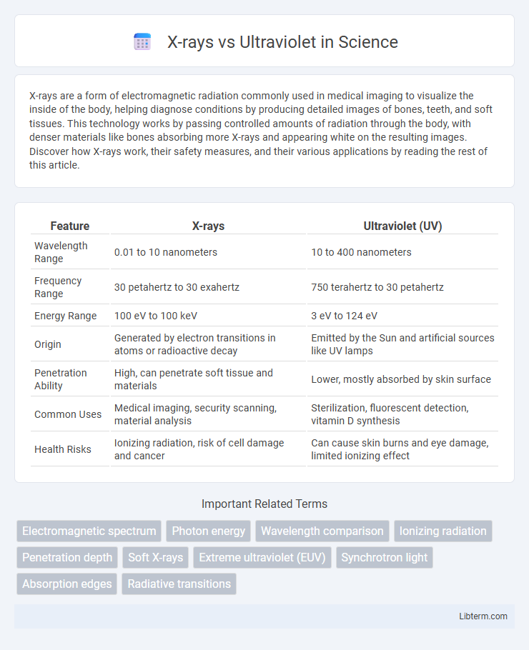
| Feature | X-rays | Ultraviolet (UV) |
|---|---|---|
| Wavelength Range | 0.01 to 10 nanometers | 10 to 400 nanometers |
| Frequency Range | 30 petahertz to 30 exahertz | 750 terahertz to 30 petahertz |
| Energy Range | 100 eV to 100 keV | 3 eV to 124 eV |
| Origin | Generated by electron transitions in atoms or radioactive decay | Emitted by the Sun and artificial sources like UV lamps |
| Penetration Ability | High, can penetrate soft tissue and materials | Lower, mostly absorbed by skin surface |
| Common Uses | Medical imaging, security scanning, material analysis | Sterilization, fluorescent detection, vitamin D synthesis |
| Health Risks | Ionizing radiation, risk of cell damage and cancer | Can cause skin burns and eye damage, limited ionizing effect |
Introduction to X-rays and Ultraviolet Radiation
X-rays and ultraviolet (UV) radiation are both forms of electromagnetic radiation with wavelengths shorter than visible light, but X-rays have much higher energy and shorter wavelengths ranging from 0.01 to 10 nanometers, while ultraviolet radiation spans wavelengths from approximately 10 to 400 nanometers. X-rays are primarily utilized in medical imaging and material analysis due to their ability to penetrate various substances, whereas ultraviolet radiation is key in processes like sterilization, fluorescence, and vitamin D synthesis. Understanding the fundamental differences in wavelength and energy between these two types of radiation is essential for their applications in science and technology.
Electromagnetic Spectrum: X-rays vs Ultraviolet Placement
X-rays occupy the electromagnetic spectrum with wavelengths ranging from 0.01 to 10 nanometers, positioned just beyond the ultraviolet region, which spans approximately 10 to 400 nanometers. Due to their shorter wavelengths and higher frequencies, X-rays possess greater energy than ultraviolet rays, enabling deeper penetration and applications in medical imaging and material analysis. The precise placement of X-rays and ultraviolet light within the electromagnetic spectrum determines their distinct interactions with matter, influencing their use in scientific, medical, and industrial fields.
Physical Properties Compared
X-rays have shorter wavelengths ranging from 0.01 to 10 nanometers, while ultraviolet (UV) rays have longer wavelengths between 10 to 400 nanometers, placing X-rays higher in the electromagnetic spectrum's frequency and energy range. X-rays possess higher photon energies, typically from 100 eV to several keV, enabling deeper penetration into materials compared to UV rays, which have energies between 3 eV and 124 eV. The electromagnetic properties of X-rays allow them to ionize atoms more effectively and pass through soft tissues and metals, whereas ultraviolet rays primarily affect surface-level molecules due to their lower energy and less penetrative capability.
Sources of X-rays and Ultraviolet Light
X-rays are primarily generated by high-energy processes such as electron collisions in X-ray tubes and astrophysical sources like black holes and neutron stars. Ultraviolet (UV) light originates from the sun, specialized bulbs, and certain chemical reactions producing emissions in the UV spectrum. Both X-rays and UV light are forms of electromagnetic radiation but differ significantly in energy levels and source mechanisms.
Penetrating Power and Wavelength Differences
X-rays possess higher penetrating power than ultraviolet rays due to their shorter wavelengths, typically ranging from 0.01 to 10 nanometers compared to ultraviolet's 10 to 400 nanometers. This shorter wavelength enables X-rays to pass through dense materials such as bone and metal, whereas ultraviolet rays are mostly absorbed by the skin and atmospheric ozone. The significant difference in energy levels, with X-rays having much higher photon energy, directly impacts their applications in medical imaging and material analysis versus ultraviolet's use in sterilization and fluorescence.
Medical and Industrial Applications
X-rays, with their high energy and penetrating ability, are extensively used in medical imaging for diagnosing fractures, dental issues, and detecting tumors, as well as in industrial non-destructive testing to inspect welds and structural integrity of materials. Ultraviolet (UV) radiation plays a crucial role in sterilization and disinfection processes within medical settings, targeting microorganisms on surfaces and in water treatment, while industrial applications include curing inks and resins in manufacturing. Both X-rays and UV light contribute uniquely to healthcare and industry, leveraging their distinct wavelengths and interaction with matter for tailored applications.
Biological Effects and Safety Concerns
X-rays possess higher energy levels than ultraviolet rays, enabling them to penetrate deeper into biological tissues and potentially cause ionization that damages DNA and increases cancer risk. Ultraviolet radiation primarily affects the skin, causing sunburn, premature aging, and DNA mutations linked to skin cancer through superficial exposure. Safety measures for X-rays involve minimizing exposure and using lead shielding, while UV protection emphasizes sunscreen, protective clothing, and limiting sun exposure to reduce harmful biological effects.
Detection and Measurement Techniques
X-rays are detected and measured using scintillation counters, Geiger-Muller tubes, and semiconductor detectors that convert high-energy photons into electrical signals. Ultraviolet radiation is measured with photodiodes, UV-sensitive photomultiplier tubes, and spectrophotometers designed to capture lower-energy UV photons. Both detection techniques rely on precise calibration and spectral sensitivity to accurately quantify radiation intensity and energy levels.
Environmental Impact and Natural Occurrence
X-rays, emitted primarily by celestial sources and human-made technologies, have limited natural environmental impact due to their low atmospheric penetration and rapid absorption. Ultraviolet (UV) radiation, predominantly produced by the sun, plays a crucial role in atmospheric chemistry, influencing ozone formation and degradation, which directly affects Earth's climate and life. While X-rays contribute minimally to environmental ionization, UV radiation drives photochemical reactions critical to ecosystems but also presents risks of biological damage and ecosystem disruption with increased exposure.
Summary: Key Differences and Similarities
X-rays and ultraviolet (UV) rays are both forms of electromagnetic radiation but differ significantly in wavelength and energy, with X-rays having much shorter wavelengths (0.01 to 10 nanometers) and higher energy than UV rays (10 to 400 nanometers). Both can cause ionization, but X-rays possess greater penetrating power, making them essential in medical imaging, while UV rays primarily affect the skin and eyes, contributing to vitamin D synthesis and potential damage like sunburn. Despite their differences, both interact with matter by increasing energy levels in atoms and molecules, influencing applications in science, medicine, and industry.
X-rays Infographic