A hematoma is a localized collection of blood outside the blood vessels, usually caused by an injury or trauma that leads to blood vessel rupture and blood pooling in tissues. Symptoms often include swelling, pain, and discoloration, and while most hematomas resolve naturally, some may require medical intervention to prevent complications. Discover more about identifying, treating, and preventing hematomas in the following article to protect your health.
Table of Comparison
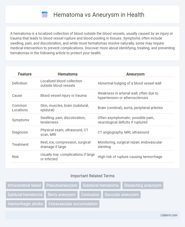
| Feature | Hematoma | Aneurysm |
|---|---|---|
| Definition | Localized blood collection outside blood vessels | Abnormal bulging of a blood vessel wall |
| Cause | Blood vessel injury or trauma | Weakness in arterial wall, often due to hypertension or atherosclerosis |
| Common Locations | Skin, muscles, brain (subdural, epidural) | Brain (cerebral), aorta, peripheral arteries |
| Symptoms | Swelling, pain, discoloration, tenderness | Often asymptomatic; possible pain, neurological deficits if ruptured |
| Diagnosis | Physical exam, ultrasound, CT scan, MRI | CT angiography, MRI, ultrasound |
| Treatment | Rest, ice, compression, surgical drainage if large | Monitoring, surgical repair, endovascular stenting |
| Risk | Usually low; complications if large or infected | High risk of rupture causing hemorrhage |
Introduction to Hematoma and Aneurysm
Hematoma is a localized collection of blood outside blood vessels, often caused by trauma or injury, resulting in swelling and discoloration. Aneurysm is a weakened, bulging area in the wall of an artery, which can expand and risk rupture, commonly occurring in the aorta or brain arteries. Both conditions require medical evaluation due to their potential to cause severe complications like bleeding or stroke.
Defining Hematoma: Causes and Types
Hematoma refers to a localized collection of blood outside blood vessels, commonly caused by trauma, surgery, or blood clotting disorders. Types of hematomas include subdural, epidural, intracerebral, and subcutaneous, each varying by location and severity. Understanding the cause and classification of hematomas aids in accurate diagnosis and effective treatment to prevent complications.
Understanding Aneurysm: Types and Risk Factors
An aneurysm is a localized bulge in the wall of a blood vessel, commonly occurring in arteries such as the aorta, brain arteries, or the peripheral vessels. Types of aneurysms include saccular, fusiform, and dissecting, each varying in shape and risk profile, with cerebral aneurysms posing high risks of rupture and hemorrhage. Risk factors for aneurysm formation include hypertension, smoking, genetic predisposition, advanced age, and connective tissue disorders like Marfan syndrome.
Key Differences: Hematoma vs Aneurysm
A hematoma is a localized collection of blood outside the blood vessels, usually caused by trauma or injury, while an aneurysm is a weakened area of a blood vessel wall that bulges or balloons, potentially leading to rupture. Hematomas often result in swelling and pain due to blood pooling in tissues, whereas aneurysms may remain asymptomatic until rupture causes severe bleeding or stroke. Diagnosis involves imaging techniques like CT or MRI scans to differentiate between the blood accumulation in hematomas and the vascular dilation characteristic of aneurysms.
Symptoms: Recognizing Hematoma and Aneurysm
Hematoma symptoms often include localized swelling, pain, discoloration, and tenderness at the injury site, sometimes accompanied by numbness or limited movement. Aneurysm symptoms vary based on location but commonly present as a pulsating lump, severe headache, vision changes, or sudden intense pain, especially if rupture occurs. Early recognition of these symptoms is critical for prompt diagnosis and treatment to prevent complications such as tissue damage from hematomas or life-threatening hemorrhage from aneurysms.
Diagnostic Methods for Hematoma and Aneurysm
Diagnostic methods for hematoma primarily include ultrasound, computed tomography (CT) scans, and magnetic resonance imaging (MRI), which help in identifying the size, location, and extent of blood accumulation. Aneurysms are typically diagnosed using CT angiography, magnetic resonance angiography (MRA), and digital subtraction angiography (DSA) to visualize arterial dilation and detect potential rupture risks. Early and accurate imaging plays a crucial role in differentiating hematomas from aneurysms and guiding appropriate treatment strategies.
Treatment Options: Hematoma Management
Hematoma management primarily involves controlling bleeding and reducing pressure on surrounding tissues, with treatment varying based on size and location. Small hematomas may resolve with rest, ice application, and compression, while larger or symptomatic hematomas often require surgical evacuation to prevent complications such as tissue necrosis or infection. Monitoring hematoma resolution through imaging techniques like ultrasound or CT scans is essential to guide treatment efficacy and adjust interventions accordingly.
Treatment Strategies for Aneurysm
Treatment strategies for aneurysm primarily include endovascular coiling and surgical clipping, which aim to prevent rupture and subsequent hemorrhage. Endovascular coiling is minimally invasive, utilizing coils to induce clotting within the aneurysm sac, effectively sealing it off from blood flow. Surgical clipping involves placing a metal clip at the aneurysm's neck to stop blood circulation, often recommended for accessible aneurysms with a high risk of rupture.
Complications and Prognosis
Hematomas can cause pressure on surrounding tissues, leading to complications such as nerve damage, tissue necrosis, or infection, with prognosis depending on the size and location of the hematoma. Aneurysms pose a significant risk of rupture, resulting in life-threatening hemorrhage, stroke, or organ damage, with prognosis influenced by the aneurysm's size, location, and promptness of medical intervention. Both conditions require timely diagnosis and treatment to minimize morbidity and improve patient outcomes.
Prevention and Long-Term Care
Preventing hematomas involves avoiding trauma, managing blood-thinning medications carefully, and controlling high blood pressure to reduce vascular injury risk. Aneurysm prevention focuses on controlling hypertension, quitting smoking, and maintaining healthy cholesterol levels to prevent arterial wall weakening. Long-term care for both conditions requires regular medical monitoring, lifestyle modifications such as dietary improvements and exercise, and adherence to prescribed treatments to minimize complications and recurrence.
Hematoma Infographic