Dacryocystitis is an infection of the tear sac causing pain, redness, and swelling near the inner corner of the eye, while dacryoadenitis affects the lacrimal gland, leading to swelling and tenderness on the outer upper eyelid. Both conditions can result in excessive tearing and discharge, requiring prompt medical attention to prevent complications and preserve eye health. Learn more about the causes, symptoms, and treatment options for your eye discomfort in the full article.
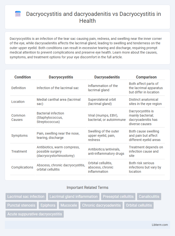
Table of Comparison
| Condition | Dacryocystitis | Dacryoadenitis | Comparison |
|---|---|---|---|
| Definition | Infection of the lacrimal sac | Inflammation of the lacrimal gland | Both affect parts of the lacrimal apparatus but differ in location |
| Location | Medial canthal area (lacrimal sac) | Superolateral orbit (lacrimal gland) | Distinct anatomical sites in the eye region |
| Common Causes | Bacterial infection (Staphylococcus, Streptococcus) | Viral (mumps, EBV), bacterial, or autoimmune | Dacryocystitis is mainly bacterial; dacryoadenitis has diverse causes |
| Symptoms | Pain, swelling near the nose, tearing, discharge | Swelling of the outer upper eyelid, pain, redness | Both cause swelling and pain but affect different eyelid areas |
| Treatment | Antibiotics, warm compress, possible surgery (dacryocystorhinostomy) | Antibiotics/antivirals, anti-inflammatory drugs | Treatment depends on infection cause and site |
| Complications | Abscess, chronic dacryocystitis, orbital cellulitis | Orbital cellulitis, abscess, chronic inflammation | Both risk serious infections but vary by location |
Introduction to Dacryocystitis and Dacryoadenitis
Dacryocystitis is an inflammation of the lacrimal sac typically caused by bacterial infection leading to pain, swelling, and tearing. Dacryoadenitis involves inflammation of the lacrimal gland, often due to viral or bacterial infections, presenting with eyelid swelling and discomfort. Differentiating between dacryocystitis and dacryoadenitis is essential for targeted treatment, as both affect different parts of the lacrimal system with distinct clinical features.
Anatomy of the Lacrimal Apparatus
Dacryocystitis primarily affects the lacrimal sac located at the medial canthus, where obstruction of the nasolacrimal duct leads to infection and inflammation. Dacryoadenitis involves inflammation of the lacrimal gland in the superolateral orbit responsible for tear production. Understanding the distinct anatomical locations--the lacrimal sac in dacryocystitis versus the lacrimal gland in dacryoadenitis--clarifies differences in pathophysiology and clinical presentation within the lacrimal apparatus.
Defining Dacryocystitis: Causes and Symptoms
Dacryocystitis is an inflammation of the lacrimal sac caused by bacterial infection, often resulting from nasolacrimal duct obstruction. Symptoms include pain, redness, and swelling near the inner corner of the eye, excessive tearing, and sometimes discharge. Unlike dacryoadenitis, which affects the lacrimal gland, dacryocystitis specifically involves the lacrimal sac, highlighting a distinct anatomical and pathological difference.
Understanding Dacryoadenitis: Etiology and Manifestations
Dacryoadenitis is an inflammatory condition affecting the lacrimal gland, often caused by viral infections like mumps or Epstein-Barr virus, bacterial infections, or autoimmune diseases such as sarcoidosis and Sjogren's syndrome. Clinical manifestations include painful swelling in the upper outer eyelid, redness, and sometimes systemic symptoms like fever, distinguishing it from dacryocystitis, which primarily involves inflammation of the lacrimal sac with symptoms localized to the inner canthus. Accurate diagnosis through clinical examination and imaging is crucial for effective treatment, which varies depending on the underlying etiology, including antibiotics for bacterial causes or corticosteroids for autoimmune-related inflammation.
Key Differences Between Dacryocystitis and Dacryoadenitis
Dacryocystitis is an infection of the lacrimal sac, often caused by obstruction in the nasolacrimal duct, leading to pain, swelling, and discharge near the inner corner of the eye. In contrast, dacryoadenitis involves inflammation of the lacrimal gland, situated in the upper outer region of the orbit, resulting in swelling of the upper eyelid and tenderness over the lacrimal gland. The key differences include the anatomical sites affected--lacrimal sac in dacryocystitis versus lacrimal gland in dacryoadenitis--and variations in clinical presentation and underlying etiology.
Risk Factors for Lacrimal Infections
Dacryocystitis and dacryoadenitis are both infections of the lacrimal apparatus but involve distinct anatomical sites; dacryocystitis affects the lacrimal sac, while dacryoadenitis targets the lacrimal gland. Risk factors for lacrimal infections include nasolacrimal duct obstruction, chronic sinusitis, trauma, and immunocompromised states, which facilitate bacterial colonization and inflammation. Understanding these factors aids in differentiating infection mechanisms and guiding targeted treatments for effective management.
Diagnostic Approaches: Dacryocystitis vs Dacryoadenitis
Diagnostic approaches for dacryocystitis primarily involve clinical examination highlighting swelling and tenderness at the lacrimal sac, often supplemented by dacryocystography or lacrimal irrigation to confirm obstruction of the nasolacrimal duct. In contrast, dacryoadenitis diagnosis emphasizes clinical signs of inflammation localized to the lacrimal gland in the superolateral orbit, supported by imaging techniques such as orbital ultrasound or CT scans to differentiate from orbital cellulitis or tumors. Both conditions may require microbial cultures from discharge or biopsy in chronic or atypical cases for accurate identification and treatment planning.
Treatment Strategies for Dacryocystitis and Dacryoadenitis
Treatment strategies for dacryocystitis focus on systemic and topical antibiotics targeting common pathogens like Staphylococcus aureus and Streptococcus species, often combined with warm compresses to facilitate drainage. Dacryoadenitis treatment involves antibiotics tailored to the underlying cause--viral or bacterial--in addition to corticosteroids for inflammatory or autoimmune etiologies when appropriate. Surgical intervention, such as dacryocystorhinostomy, is reserved for chronic or refractory dacryocystitis, while dacryoadenitis rarely requires surgery unless abscess formation occurs.
Prognosis and Complications
Dacryocystitis typically has a favorable prognosis with prompt antibiotic therapy, though delayed treatment can lead to complications like lacrimal sac abscess or chronic dacryocystitis causing persistent epiphora. Dacryoadenitis often resolves with appropriate antimicrobial or anti-inflammatory treatment, but may result in orbital cellulitis or abscess if untreated. Compared to dacryocystitis, dacryoadenitis poses a higher risk for complications involving the lacrimal gland and adjacent orbital structures, potentially leading to more severe ocular morbidity.
Prevention and Patient Education
Effective prevention of dacryocystitis involves maintaining proper eye hygiene and prompt treatment of nasolacrimal duct obstruction to reduce infection risk. Educating patients on recognizing early symptoms such as swelling and pain near the tear sac encourages timely medical intervention, minimizing complications. In contrast, prevention of dacryoadenitis centers on avoiding systemic infections and managing autoimmune conditions, while patient education emphasizes recognizing symptoms related to lacrimal gland inflammation.
Dacryocystitis and dacryoadenitis Infographic