Myocardial relaxation is a critical phase in the cardiac cycle where heart muscle fibers return to their resting state after contraction, allowing the chambers to fill with blood efficiently. Proper relaxation ensures optimal blood flow and maintains healthy cardiac output, preventing conditions like diastolic dysfunction and heart failure. Discover how understanding myocardial relaxation can improve your heart health and the implications for diagnosing and treating cardiac diseases in the rest of this article.
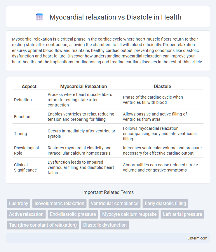
Table of Comparison
| Aspect | Myocardial Relaxation | Diastole |
|---|---|---|
| Definition | Process where heart muscle fibers return to resting state after contraction | Phase of the cardiac cycle when ventricles fill with blood |
| Function | Enables ventricles to relax, reducing tension and preparing for filling | Allows passive and active filling of ventricles from atria |
| Timing | Occurs immediately after ventricular systole | Follows myocardial relaxation, encompassing early and late ventricular filling |
| Physiological Role | Restores myocardial elasticity and intracellular calcium homeostasis | Increases ventricular volume and pressure necessary for effective cardiac output |
| Clinical Significance | Dysfunction leads to impaired ventricular filling and diastolic heart failure | Abnormalities can cause reduced stroke volume and congestive symptoms |
Introduction to Myocardial Relaxation and Diastole
Myocardial relaxation refers to the phase during which the heart muscle relaxes after contraction, allowing the chambers to expand and fill with blood. Diastole represents this relaxation period in the cardiac cycle, encompassing both the relaxation of myocardial fibers and ventricular filling. Understanding the distinction between active myocardial relaxation and the mechanical events of diastole is crucial for assessing cardiac function and diagnosing diastolic dysfunction.
Defining Myocardial Relaxation
Myocardial relaxation refers to the active biochemical process during which cardiac muscle fibers decrease tension after contraction, enabling ventricular chambers to expand and fill with blood. It is a critical phase within diastole, specifically encompassing early diastole, where calcium ions are reabsorbed into the sarcoplasmic reticulum, leading to muscle relaxation and ventricular filling. Efficient myocardial relaxation ensures optimal diastolic function and cardiac output, distinguishing it from the broader diastolic phase that includes both relaxation and passive ventricular filling.
Understanding Cardiac Diastole
Myocardial relaxation refers specifically to the process of the heart muscle fibers ceasing contraction and returning to their resting state, which is a critical component of cardiac diastole. Cardiac diastole encompasses the entire phase of the heartbeat when the heart chambers fill with blood, including both isovolumetric relaxation and ventricular filling phases. Effective myocardial relaxation ensures optimal ventricular filling and contributes significantly to maintaining adequate stroke volume and cardiac output during diastole.
Key Differences Between Myocardial Relaxation and Diastole
Myocardial relaxation refers specifically to the active biochemical process where cardiac muscle fibers return to their resting state following contraction, involving calcium reuptake and muscle fiber elongation. Diastole encompasses the broader phase of the cardiac cycle including myocardial relaxation, characterized by ventricular filling and chamber expansion. Key differences include that myocardial relaxation is a cellular event within the myocardium, while diastole represents the mechanical and hemodynamic phase impacting overall cardiac function and blood flow.
The Physiology of Ventricular Relaxation
Myocardial relaxation refers specifically to the active biochemical and mechanical process by which ventricular muscle fibers cease contraction and restore resting tension, crucially involving calcium ion reuptake into the sarcoplasmic reticulum. Diastole encompasses the entire phase of the cardiac cycle when the ventricles fill with blood, including isovolumetric relaxation and ventricular filling. Efficient ventricular relaxation ensures optimal diastolic function by enabling adequate ventricular compliance and low end-diastolic pressure, essential for effective cardiac output and coronary perfusion.
Molecular Mechanisms of Myocardial Relaxation
Myocardial relaxation primarily involves calcium reuptake into the sarcoplasmic reticulum through SERCA2a activity, regulated by phospholamban phosphorylation, which decreases cytosolic calcium, facilitating cardiac muscle fiber relaxation. During diastole, this molecular process ensures effective ventricular filling by reducing myocardial tension and promoting chamber compliance. Dysregulation of these calcium-handling proteins contributes to impaired relaxation seen in diastolic dysfunction and heart failure with preserved ejection fraction.
Clinical Assessment of Diastolic Function
Myocardial relaxation is a crucial active process during diastole, enabling efficient ventricular filling and maintaining cardiac output. Clinical assessment of diastolic function relies on echocardiographic parameters such as E/A ratio, E/e' ratio, deceleration time, and left atrial volume to evaluate impaired relaxation, filling pressures, and compliance. Accurate interpretation of these measurements helps diagnose diastolic dysfunction, guiding management of heart failure with preserved ejection fraction (HFpEF).
Impact of Myocardial Relaxation on Cardiac Health
Myocardial relaxation, a critical phase within diastole, directly influences left ventricular filling and overall cardiac output by enabling efficient blood inflow. Impaired myocardial relaxation leads to diastolic dysfunction, increasing the risk of heart failure with preserved ejection fraction (HFpEF) and elevated cardiac filling pressures. Optimizing myocardial relaxation through targeted therapies enhances cardiac performance and reduces morbidity associated with diastolic heart disease.
Disorders Involving Impaired Relaxation and Diastole
Impaired myocardial relaxation primarily affects diastolic function, leading to conditions such as diastolic dysfunction and heart failure with preserved ejection fraction (HFpEF). Disorders like hypertrophic cardiomyopathy and restrictive cardiomyopathy cause stiffness of the ventricular walls, reducing compliance and hindering proper ventricular filling during diastole. Elevated left atrial pressure and pulmonary congestion often result from this impaired relaxation, highlighting the clinical significance of diastolic abnormalities.
Summary: Importance in Cardiology Practice
Myocardial relaxation is the active process of ventricular muscle fibers lengthening and reducing tension after contraction, crucial for effective ventricular filling during diastole. Diastole refers to the phase of the cardiac cycle when the heart chambers relax and fill with blood, directly influenced by the quality of myocardial relaxation. Accurate assessment of myocardial relaxation and diastolic function is vital in cardiology for diagnosing heart failure with preserved ejection fraction and guiding appropriate treatment strategies.
Myocardial relaxation Infographic