Enophthalmos occurs when the eye sinks deeper into the orbit, often due to trauma, orbital fractures, or certain medical conditions affecting the orbital fat or muscles. Accurate diagnosis involves clinical examination and imaging techniques such as CT or MRI to identify underlying causes and guide appropriate treatment. Discover how understanding enophthalmos can improve your approach to diagnosis and management in the full article.
Table of Comparison
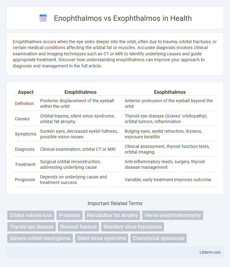
| Aspect | Enophthalmos | Exophthalmos |
|---|---|---|
| Definition | Posterior displacement of the eyeball within the orbit | Anterior protrusion of the eyeball beyond the orbit |
| Causes | Orbital trauma, silent sinus syndrome, orbital fat atrophy | Thyroid eye disease (Graves' orbitopathy), orbital tumors, inflammation |
| Symptoms | Sunken eyes, decreased eyelid fullness, possible vision issues | Bulging eyes, eyelid retraction, dryness, exposure keratitis |
| Diagnosis | Clinical examination, orbital CT or MRI | Clinical assessment, thyroid function tests, orbital imaging |
| Treatment | Surgical orbital reconstruction, addressing underlying cause | Anti-inflammatory meds, surgery, thyroid disease management |
| Prognosis | Depends on underlying cause and treatment success | Variable; early treatment improves outcome |
Understanding Enophthalmos and Exophthalmos
Enophthalmos is the posterior displacement of the eyeball within the orbit, often caused by trauma, orbital fractures, or fat atrophy, leading to a sunken eye appearance. Exophthalmos, also known as proptosis, involves the forward protrusion of the eyeball, commonly associated with thyroid eye disease, tumors, or inflammation, resulting in bulging eyes. Differentiating between enophthalmos and exophthalmos is critical for diagnosing underlying conditions and guiding appropriate treatment in ophthalmology.
Key Differences Between Enophthalmos and Exophthalmos
Enophthalmos is characterized by the posterior displacement of the eyeball within the orbit, often caused by trauma, fat atrophy, or orbital fractures. Exophthalmos involves the anterior protrusion of the eyeball, commonly associated with thyroid eye disease or orbital tumors. The key difference lies in the direction of eyeball displacement, with enophthalmos leading to sunken eyes and exophthalmos resulting in bulging eyes.
Causes of Enophthalmos
Enophthalmos is primarily caused by trauma leading to orbital fractures, fat atrophy or loss, and conditions such as silent sinus syndrome where the maxillary sinus collapses, pulling the eye inward. Other causes include chronic inflammation or fibrosis from diseases like scleroderma, and orbital tumors that reduce orbital volume. Understanding these underlying causes is critical for accurate diagnosis and treatment to differentiate from exophthalmos, which involves protrusion of the eye and is often related to thyroid eye disease or orbital masses.
Causes of Exophthalmos
Exophthalmos, characterized by the abnormal protrusion of the eyeball, is primarily caused by Graves' disease, an autoimmune disorder leading to inflammation and swelling of the orbital tissues. Other causes include orbital tumors, infections such as orbital cellulitis, and trauma that result in increased orbital volume or pressure. In contrast, enophthalmos involves the posterior displacement of the eye, often due to orbital fractures, fat atrophy, or chronic sinus disease.
Clinical Signs and Symptoms
Enophthalmos presents with posterior displacement of the eyeball within the orbit, causing a sunken appearance, often accompanied by diplopia and enophthalmic furrows. Exophthalmos is characterized by anterior protrusion of the eyeball, commonly seen in thyroid eye disease, with symptoms such as eyelid retraction, conjunctival injection, and impaired ocular motility. Both conditions may lead to visual disturbances but differ primarily in globe position and associated orbital pathology.
Diagnostic Methods for Orbital Disorders
Diagnostic methods for orbital disorders differentiate enophthalmos and exophthalmos primarily through imaging techniques such as computed tomography (CT) and magnetic resonance imaging (MRI), which assess orbital volume changes and soft tissue abnormalities. Hertel exophthalmometry provides quantitative measurement of globe protrusion or retraction, aiding in the clinical evaluation of proptosis or enophthalmos. Ultrasonography and orbital ultrasound Doppler also assist in evaluating orbital mass lesions or vascular abnormalities contributing to these conditions.
Complications Associated with Enophthalmos and Exophthalmos
Enophthalmos, characterized by posterior displacement of the eyeball, can lead to complications such as diplopia, orbital pain, and impaired ocular motility due to orbital fat atrophy or trauma. Exophthalmos, or proptosis, increases the risk of exposure keratopathy, corneal ulceration, and optic neuropathy caused by increased orbital pressure or thyroid eye disease. Both conditions require careful monitoring to prevent vision loss and manage underlying etiologies effectively.
Treatment Options and Management Strategies
Enophthalmos treatment involves addressing underlying causes such as trauma, orbital fractures, or fat atrophy, often requiring orbital reconstruction or volume augmentation with fillers or implants. Exophthalmos management focuses on reducing proptosis typically due to thyroid eye disease, with options including corticosteroids, orbital decompression surgery, radiotherapy, and eyelid surgery to protect the cornea and improve ocular function. Both conditions benefit from multidisciplinary approaches involving ophthalmologists, endocrinologists, and maxillofacial surgeons to optimize functional and cosmetic outcomes.
Prognosis and Long-term Outcomes
Enophthalmos, characterized by the posterior displacement of the eye within the orbit, often results from trauma or orbital fat atrophy, with a prognosis dependent on timely intervention and severity of underlying cause. Exophthalmos, commonly associated with thyroid eye disease, presents as protrusion of the eyeball and requires management to prevent complications like exposure keratopathy or optic neuropathy, with long-term outcomes varying based on disease control and treatment efficacy. Both conditions demand specialized ophthalmic evaluation to optimize functional and cosmetic results, impacting patients' quality of life differently.
Prevention and Patient Education
Preventing enophthalmos involves managing underlying causes such as trauma, orbital tumors, or chronic sinus infections through timely medical intervention and protective measures like wearing safety gear during high-risk activities. Patient education emphasizes recognizing early symptoms, seeking prompt diagnosis, and adhering to treatment plans to prevent progression or complications, especially in cases of enophthalmos caused by orbital fat atrophy or fractures. For exophthalmos, common in thyroid eye disease, prevention includes controlling thyroid function with medications, regular ophthalmologic evaluations, and lifestyle modifications to reduce inflammation and eye irritation.
Enophthalmos Infographic