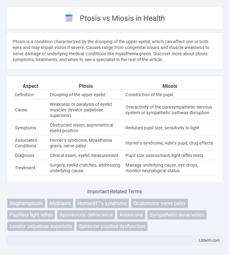
Ptosis is a condition characterized by the drooping of the upper eyelid, which can affect one or both eyes and may impair vision if severe. Causes range from congenital issues and muscle weakness to nerve damage or underlying medical conditions like myasthenia gravis. Discover more about ptosis symptoms, treatments, and when to see a specialist in the rest of the article.
Table of Comparison
| Aspect | Ptosis | Miosis |
|---|---|---|
| Definition | Drooping of the upper eyelid | Constriction of the pupil |
| Cause | Weakness or paralysis of eyelid muscles (levator palpebrae superioris) | Overactivity of the parasympathetic nervous system or sympathetic pathway disruption |
| Symptoms | Obstructed vision, asymmetrical eyelid position | Reduced pupil size, sensitivity to light |
| Associated Conditions | Horner's syndrome, Myasthenia gravis, nerve palsy | Horner's syndrome, Adie's pupil, drug effects |
| Diagnosis | Clinical exam, eyelid measurement | Pupil size assessment, light reflex tests |
| Treatment | Surgery, eyelid crutches, addressing underlying cause | Manage underlying cause, eye drops, monitor neurological status |
Introduction to Ptosis and Miosis
Ptosis refers to the drooping of the upper eyelid caused by muscle weakness, nerve damage, or underlying medical conditions such as Horner's syndrome or myasthenia gravis. Miosis is the abnormal constriction of the pupil often resulting from parasympathetic nerve stimulation, opioid use, or brainstem injury. Both symptoms can indicate neurological disorders and require thorough clinical evaluation to determine the underlying cause.
Defining Ptosis: Causes and Symptoms
Ptosis, characterized by the drooping of the upper eyelid, results from dysfunction of the levator muscle or its nerve supply, commonly caused by factors such as nerve damage, muscle diseases, or aging. Symptoms include a visibly lowered eyelid that may obstruct vision, eye fatigue, and sometimes asymmetry between the eyes. Distinct from miosis, which refers to pupil constriction, ptosis specifically affects eyelid position and function.
Understanding Miosis: Causes and Symptoms
Miosis, characterized by abnormal constriction of the pupil, can result from factors such as exposure to opioids, brainstem injuries, or Horner's syndrome. Unlike ptosis, which involves drooping of the eyelid, miosis primarily affects pupil size and light response. Recognizing symptoms like pinpoint pupils and reduced reaction to light helps differentiate miosis and identify underlying neurological or pharmacological causes.
Key Differences Between Ptosis and Miosis
Ptosis is the drooping of the upper eyelid caused by muscle weakness or nerve damage, while miosis refers to the abnormal constriction of the pupil due to parasympathetic nervous system activation. Ptosis primarily affects eyelid position and can impair vision, whereas miosis impacts pupil size, affecting the amount of light entering the eye and vision clarity. Both conditions may coexist in Horner's syndrome, highlighting their distinct neurological origins but overlapping clinical presentation.
Common Conditions Associated with Ptosis
Ptosis, characterized by the drooping of the upper eyelid, is commonly associated with conditions such as Horner's syndrome, myasthenia gravis, and oculomotor nerve palsy. Unlike miosis, which involves pupillary constriction often seen in Horner's syndrome or opioid use, ptosis specifically indicates muscular or neurological impairment affecting eyelid elevation. Accurate diagnosis requires evaluating underlying causes like neuromuscular disorders, traumatic injury, or congenital abnormalities to differentiate ptosis from other ophthalmic signs.
Disorders Linked to Miosis
Disorders linked to miosis primarily include Horner's syndrome, characterized by ptosis, miosis, and anhidrosis due to sympathetic pathway disruption, and opioid intoxication, where excessive parasympathetic stimulation causes pinpoint pupils. Other neurological conditions such as brainstem strokes and pontine hemorrhages can lead to miosis by affecting the Edinger-Westphal nucleus. Chemical exposure to organophosphates also induces miosis through acetylcholinesterase inhibition, resulting in excessive acetylcholine accumulation.
Diagnostic Approaches for Ptosis and Miosis
Diagnostic approaches for ptosis involve detailed clinical examination to assess eyelid drooping severity, including measuring the margin-reflex distance and levator function tests. Miosis diagnosis requires pupillary size evaluation under various lighting conditions and pharmacologic testing to determine if the constriction is due to parasympathetic overactivity or sympathetic pathway disruption. Imaging studies like MRI or CT scans support differential diagnosis when neurologic or systemic causes for ptosis and miosis, such as Horner's syndrome, are suspected.
Treatment Options for Ptosis
Ptosis treatment options depend on the severity and cause, ranging from conservative approaches like eyelid exercises and prescription glasses with a ptosis crutch to surgical interventions such as levator resection or frontalis sling procedures. In cases caused by underlying neurological conditions, addressing the primary disorder is essential before considering eyelid surgery. Non-surgical methods, including the use of special contact lenses or ptosis props, may provide temporary relief but are generally less effective than surgical correction for long-term improvement.
Management Strategies for Miosis
Management strategies for miosis primarily involve addressing the underlying cause, such as treating opioid overdose with naloxone or managing exposure to organophosphates using atropine. In cases of pharmacologic miosis, discontinuation or adjustment of causative medications like pilocarpine or brimonidine is essential. Symptomatic relief and pupil dilation may be achieved with mydriatic agents, but careful evaluation by a healthcare professional is critical to guide appropriate treatment and exclude serious neurologic conditions.
Prognosis and Outlook for Patients with Ptosis or Miosis
Prognosis for patients with ptosis varies depending on the underlying cause, with congenital ptosis often requiring surgical correction to prevent amblyopia, while acquired ptosis linked to neurological disorders may reflect the progression of the primary disease. Miosis prognosis depends largely on whether it is caused by benign factors such as pharmacologic agents or serious conditions like Horner's syndrome, where timely identification and treatment of the root cause can improve patient outcomes. Both ptosis and miosis require accurate diagnosis and targeted management to optimize visual function and address any associated systemic conditions.
Ptosis Infographic

 libterm.com