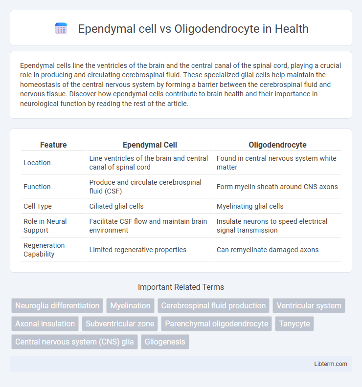
Ependymal cells line the ventricles of the brain and the central canal of the spinal cord, playing a crucial role in producing and circulating cerebrospinal fluid. These specialized glial cells help maintain the homeostasis of the central nervous system by forming a barrier between the cerebrospinal fluid and nervous tissue. Discover how ependymal cells contribute to brain health and their importance in neurological function by reading the rest of the article.
Table of Comparison
| Feature | Ependymal Cell | Oligodendrocyte |
|---|---|---|
| Location | Line ventricles of the brain and central canal of spinal cord | Found in central nervous system white matter |
| Function | Produce and circulate cerebrospinal fluid (CSF) | Form myelin sheath around CNS axons |
| Cell Type | Ciliated glial cells | Myelinating glial cells |
| Role in Neural Support | Facilitate CSF flow and maintain brain environment | Insulate neurons to speed electrical signal transmission |
| Regeneration Capability | Limited regenerative properties | Can remyelinate damaged axons |
Introduction to Glial Cells in the CNS
Ependymal cells and oligodendrocytes are essential types of glial cells in the central nervous system (CNS) with distinct functions; ependymal cells line the ventricles and produce cerebrospinal fluid, while oligodendrocytes form myelin sheaths around axons to facilitate rapid electrical signal conduction. These glial cells contribute to CNS homeostasis and neural support, with ependymal cells involved in barrier formation and cerebrospinal fluid circulation, and oligodendrocytes crucial for insulating neurons and enhancing synaptic transmission. Understanding their roles contributes to insights into neurodevelopmental processes and CNS repair mechanisms.
Overview of Ependymal Cells
Ependymal cells line the ventricles of the brain and the central canal of the spinal cord, playing a crucial role in producing and circulating cerebrospinal fluid (CSF). These ciliated glial cells form a barrier between the CSF and nervous tissue, contributing to homeostasis and nutrient transport within the central nervous system (CNS). Unlike oligodendrocytes, which are responsible for myelinating CNS axons, ependymal cells primarily facilitate CSF movement and maintain the brain's internal environment.
Overview of Oligodendrocytes
Oligodendrocytes are specialized glial cells in the central nervous system responsible for the formation and maintenance of myelin sheaths around neuronal axons, enhancing rapid electrical impulse conduction. Unlike ependymal cells, which line the brain ventricles and spinal cord central canal to produce cerebrospinal fluid, oligodendrocytes provide metabolic support to neurons and contribute to neural signal insulation. The unique multi-branching structure of oligodendrocytes enables them to myelinate multiple axon segments simultaneously, critical for efficient neural communication.
Origin and Developmental Differences
Ependymal cells originate from the neuroepithelium of the neural tube, playing a crucial role in lining the ventricular system and facilitating cerebrospinal fluid (CSF) circulation. Oligodendrocytes develop from oligodendrocyte precursor cells (OPCs) derived from the ventricular zone, responsible for myelinating axons in the central nervous system to enhance neural signal transmission. The distinct developmental pathways reflect their specialized functions, with ependymal cells maintaining CNS homeostasis and oligodendrocytes supporting neural connectivity.
Structural Characteristics and Morphology
Ependymal cells are ciliated, cuboidal to columnar epithelial cells lining the ventricles of the brain and central canal of the spinal cord, featuring microvilli that aid in cerebrospinal fluid circulation. Oligodendrocytes possess a smaller, round cell body with fewer processes that extend to form myelin sheaths around multiple axons in the central nervous system. The unique morphology of ependymal cells supports barrier and fluid movement functions, while oligodendrocytes specialize in insulation and rapid electrical conduction.
Functional Roles in the Central Nervous System
Ependymal cells line the ventricles of the brain and spinal cord, playing a crucial role in producing and circulating cerebrospinal fluid to maintain the central nervous system's homeostasis. Oligodendrocytes are responsible for forming the myelin sheath around CNS axons, facilitating rapid electrical impulse conduction and supporting neuronal integrity. Both cell types contribute fundamentally to CNS function, with ependymal cells regulating the extracellular environment and oligodendrocytes enhancing neural signal transmission.
Involvement in Neurological Disorders
Ependymal cells contribute to neurological disorders by affecting cerebrospinal fluid circulation, which can lead to hydrocephalus and impair waste clearance in neurodegenerative diseases. Oligodendrocytes are crucial in multiple sclerosis due to their role in myelin sheath formation, with demyelination causing significant neural signal disruption. Dysfunction in either cell type can exacerbate pathology in central nervous system disorders, impacting neuronal health and repair mechanisms.
Key Molecular Markers and Identification
Ependymal cells are characterized by the expression of markers such as S100b, vimentin, and CD24, which aid in their identification as ciliated cells lining the ventricles and central canal of the brain and spinal cord. Oligodendrocytes express distinct molecular markers including Olig2, myelin basic protein (MBP), and proteolipid protein (PLP), essential for their role in myelin sheath formation around CNS axons. Immunohistochemistry targeting these specific proteins enables precise differentiation between ependymal cells and oligodendrocytes in neural tissue samples.
Interactions with Other Neural Cells
Ependymal cells form a crucial interface by lining the ventricles of the brain and producing cerebrospinal fluid, facilitating nutrient exchange and waste removal that supports neurons and glial cells. Oligodendrocytes interact directly with neurons by wrapping their axons in myelin sheaths, enhancing electrical signal conduction and providing metabolic support. The collaboration between ependymal cells and oligodendrocytes ensures a stable neural environment, promoting efficient communication and homeostasis within the central nervous system.
Summary: Comparing Ependymal Cells vs Oligodendrocytes
Ependymal cells line the ventricles of the brain and the central canal of the spinal cord, playing a critical role in the production and circulation of cerebrospinal fluid (CSF). Oligodendrocytes are responsible for myelinating axons in the central nervous system, enhancing the speed and efficiency of electrical signal transmission. While ependymal cells contribute to neural environment regulation, oligodendrocytes primarily support neural function through insulation of neuronal fibers.
Ependymal cell Infographic

 libterm.com