Sialadenitis is the inflammation of the salivary glands, often caused by bacterial or viral infections, leading to painful swelling and sometimes pus formation. Ludwig's Angina is a severe and potentially life-threatening cellulitis that affects the floor of the mouth and neck, originating from infected salivary glands or dental abscesses. Discover more about the symptoms, causes, and treatment options to protect your oral health and prevent serious complications.
Table of Comparison
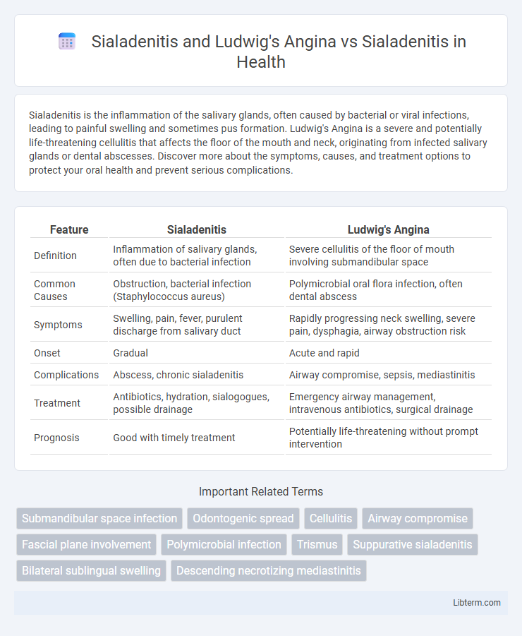
| Feature | Sialadenitis | Ludwig's Angina |
|---|---|---|
| Definition | Inflammation of salivary glands, often due to bacterial infection | Severe cellulitis of the floor of mouth involving submandibular space |
| Common Causes | Obstruction, bacterial infection (Staphylococcus aureus) | Polymicrobial oral flora infection, often dental abscess |
| Symptoms | Swelling, pain, fever, purulent discharge from salivary duct | Rapidly progressing neck swelling, severe pain, dysphagia, airway obstruction risk |
| Onset | Gradual | Acute and rapid |
| Complications | Abscess, chronic sialadenitis | Airway compromise, sepsis, mediastinitis |
| Treatment | Antibiotics, hydration, sialogogues, possible drainage | Emergency airway management, intravenous antibiotics, surgical drainage |
| Prognosis | Good with timely treatment | Potentially life-threatening without prompt intervention |
Overview of Sialadenitis
Sialadenitis is an inflammation of the salivary glands, primarily caused by bacterial or viral infection, often resulting in swelling, pain, and reduced saliva flow. Ludwig's angina is a serious, rapidly progressing cellulitis involving the submandibular space, often secondary to dental infections, which can lead to airway obstruction and requires urgent medical intervention. In contrast, simple sialadenitis typically affects the parotid or submandibular glands and is managed with antibiotics, hydration, and gland massage without the critical airway risks associated with Ludwig's angina.
Pathophysiology of Sialadenitis
Sialadenitis is an inflammatory condition of the salivary glands caused primarily by bacterial or viral infections, leading to glandular swelling, pain, and reduced saliva flow due to ductal obstruction or altered salivary composition. Ludwig's Angina, a severe cellulitis involving the submandibular and sublingual spaces, results from untreated sialadenitis or odontogenic infections, causing rapid airway compromise through diffuse inflammation and edema. The pathophysiology of sialadenitis involves ductal obstruction, retrograde bacterial invasion, and subsequent glandular inflammation, often exacerbated by factors such as dehydration, immunosuppression, or salivary stasis.
Etiology and Causes of Sialadenitis
Sialadenitis primarily results from bacterial infections, most commonly Staphylococcus aureus or Streptococcus species, causing inflammation of the salivary glands due to ductal obstruction or decreased salivary flow. Ludwig's Angina, a severe cellulitis involving the submandibular space, often arises from infected mandibular molar teeth and can lead to secondary sialadenitis due to adjacent gland involvement. Identifying the etiology of sialadenitis is critical, as it often stems from systemic factors such as dehydration, immunosuppression, or salivary gland duct stones (sialolithiasis), distinguishing it from the primarily odontogenic origins of Ludwig's Angina.
Clinical Presentation of Sialadenitis
Sialadenitis typically presents with pain and swelling in the affected salivary gland, often accompanied by fever, malaise, and purulent discharge from the duct. In contrast, Ludwig's angina manifests as rapidly progressing cellulitis of the submandibular space causing severe neck swelling, dysphagia, and potential airway obstruction. Clinical presentation of sialadenitis is localized, whereas Ludwig's angina involves extensive submandibular and sublingual tissue infection leading to systemic toxicity.
Diagnostic Modalities for Sialadenitis
Diagnostic modalities for sialadenitis primarily include ultrasound imaging, which provides detailed visualization of salivary gland inflammation and ductal obstruction, and sialography, which helps identify glandular ductal abnormalities. MRI and CT scans are utilized in complicated cases to assess the extent of infection and distinguish sialadenitis from Ludwig's angina, a condition characterized by bilateral cellulitis of the submandibular space with potential airway compromise. Laboratory tests such as elevated inflammatory markers and bacterial cultures complement imaging to confirm diagnosis and guide treatment planning.
What is Ludwig's Angina?
Ludwig's Angina is a rapidly progressing cellulitis of the submandibular, sublingual, and submental spaces, often resulting from untreated dental infections and leading to airway obstruction. Unlike sialadenitis, which is inflammation of the salivary glands typically caused by bacterial or viral infections, Ludwig's Angina involves a deeper, diffusely spreading infection affecting connective tissues under the floor of the mouth. Prompt recognition and aggressive treatment with antibiotics and airway management are critical to prevent life-threatening complications in Ludwig's Angina.
Ludwig's Angina vs Sialadenitis: Key Differences
Ludwig's angina is a rapidly progressing cellulitis involving the floor of the mouth and submandibular space, often caused by polymicrobial infections originating from dental sources, while sialadenitis refers to inflammation of the salivary glands typically due to bacterial or viral infections or obstruction. Ludwig's angina presents with severe swelling, pain, and airway compromise requiring urgent airway management, contrasted with sialadenitis which mainly manifests as localized gland swelling, tenderness, and sometimes pus drainage without immediate airway risk. The key clinical distinction lies in Ludwig's angina's potential for life-threatening airway obstruction versus sialadenitis' more localized and less emergent pathology.
Clinical Features of Ludwig's Angina
Ludwig's Angina presents with rapid onset of bilateral submandibular swelling, severe floor-of-mouth elevation, and marked trismus, often leading to airway obstruction, distinguishing it from typical sialadenitis. Patients frequently exhibit fever, dysphagia, and a "woody" induration without abscess formation, unlike the localized gland tenderness and purulent discharge seen in sialadenitis. Early recognition of these clinical features is critical to prevent life-threatening complications associated with Ludwig's Angina.
Management and Treatment Approaches
Management of sialadenitis involves antibiotics targeting Staphylococcus aureus and anaerobic bacteria, along with hydration, sialogogues, and warm compresses to promote salivary flow and gland drainage. Ludwig's angina requires aggressive airway management, intravenous broad-spectrum antibiotics covering polymicrobial flora, and often surgical intervention for abscess drainage to prevent airway obstruction. Prompt differentiation and tailored treatment are critical due to Ludwig's angina's rapid progression and potential life-threatening complications compared to the typically less urgent treatment of uncomplicated sialadenitis.
Complications and Prognosis: Sialadenitis vs Ludwig’s Angina
Sialadenitis, an inflammation of the salivary glands, can lead to abscess formation, glandular destruction, or chronic ductal obstruction if untreated, with generally favorable prognosis upon timely antibiotic therapy. Ludwig's Angina, a rapidly progressing cellulitis of the submandibular space, often results in airway obstruction, sepsis, and mediastinitis, carrying a higher risk of mortality without emergent intervention. Prognosis is significantly poorer in Ludwig's Angina due to its aggressive nature and potential for life-threatening complications compared to the more localized and manageable course of sialadenitis.
Sialadenitis and Ludwig's Angina Infographic