A mucocele is a benign cyst-like lesion caused by the accumulation of mucus, typically occurring in the oral cavity or sinuses due to blocked salivary glands or mucus ducts. These swellings can cause discomfort and sometimes interfere with speech or eating, highlighting the importance of timely diagnosis and treatment. Learn more about the causes, symptoms, and effective management options to protect your oral health in the rest of this article.
Table of Comparison
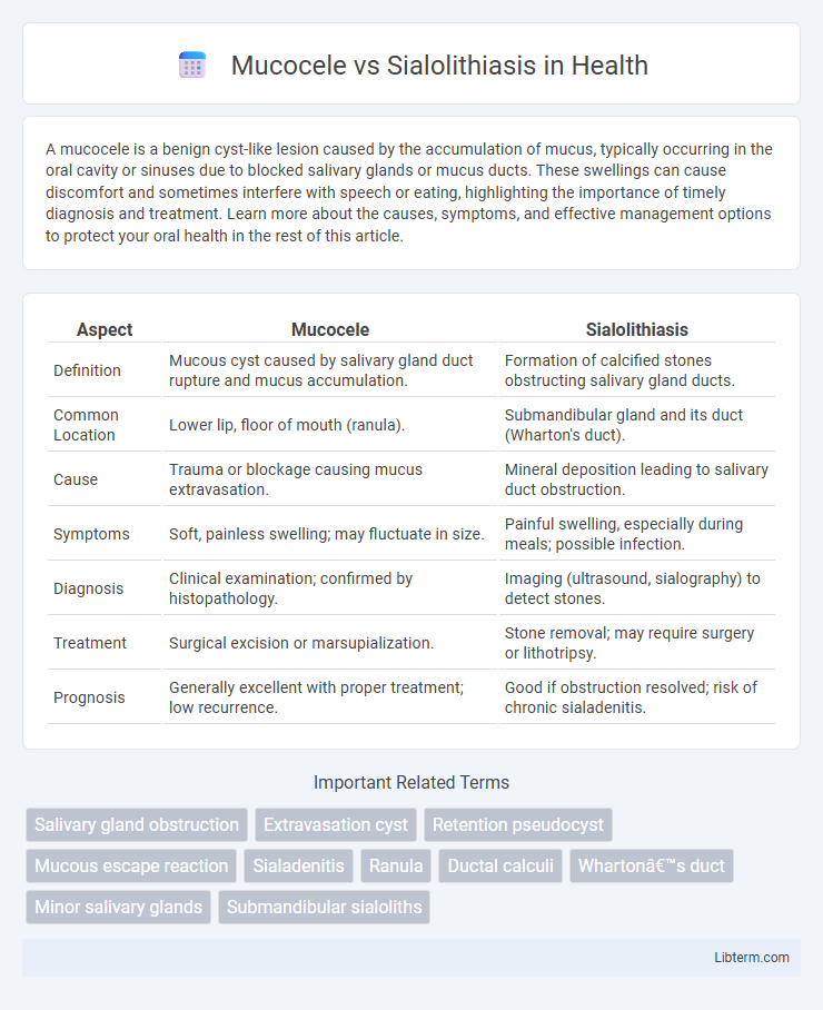
| Aspect | Mucocele | Sialolithiasis |
|---|---|---|
| Definition | Mucous cyst caused by salivary gland duct rupture and mucus accumulation. | Formation of calcified stones obstructing salivary gland ducts. |
| Common Location | Lower lip, floor of mouth (ranula). | Submandibular gland and its duct (Wharton's duct). |
| Cause | Trauma or blockage causing mucus extravasation. | Mineral deposition leading to salivary duct obstruction. |
| Symptoms | Soft, painless swelling; may fluctuate in size. | Painful swelling, especially during meals; possible infection. |
| Diagnosis | Clinical examination; confirmed by histopathology. | Imaging (ultrasound, sialography) to detect stones. |
| Treatment | Surgical excision or marsupialization. | Stone removal; may require surgery or lithotripsy. |
| Prognosis | Generally excellent with proper treatment; low recurrence. | Good if obstruction resolved; risk of chronic sialadenitis. |
Introduction to Mucocele and Sialolithiasis
Mucocele is a common benign cystic lesion of the salivary glands, typically caused by the rupture or obstruction of minor salivary gland ducts, leading to mucus accumulation in the surrounding tissues. Sialolithiasis refers to the formation of calcified concretions or salivary stones within the salivary ducts, most frequently affecting the submandibular gland, resulting in ductal obstruction and subsequent gland inflammation. Both conditions disrupt normal salivary flow but differ in etiology, clinical presentation, and management strategies.
Definition and Pathophysiology
Mucocele is a mucous cyst that forms due to the rupture or blockage of a salivary gland duct, leading to mucus accumulation in the surrounding tissues. Sialolithiasis involves the formation of calcified stones within the salivary glands or ducts, causing obstruction and inflammation. Both conditions disrupt normal salivary flow but differ in etiology: mucocele arises from ductal trauma or blockage by mucus, while sialolithiasis results from mineral precipitation forming sialoliths.
Causes and Risk Factors
Mucocele results from the rupture or blockage of salivary gland ducts, leading to mucus accumulation often triggered by trauma or lip biting. Sialolithiasis occurs due to the formation of calcified stones within the salivary glands, commonly caused by dehydration, reduced salivary flow, or altered saliva composition. Risk factors for mucocele include repetitive lip injury and duct obstruction, while sialolithiasis is associated with age, male gender, and systemic conditions like Sjogren's syndrome or chronic infections.
Clinical Presentation and Symptoms
Mucocele typically presents as a painless, bluish, fluctuant swelling on the lower lip or oral mucosa, resulting from mucus extravasation due to minor salivary gland duct rupture. Sialolithiasis manifests with recurrent pain and swelling in the affected salivary gland, especially during meals, caused by obstruction from calcified salivary stones. Both conditions can impair salivary flow but differ significantly in symptom onset, location, and associated discomfort levels.
Diagnostic Techniques and Imaging
Mucocele typically presents as a painless, translucent swelling in the oral mucosa, diagnosed primarily through clinical examination and confirmed by imaging modalities such as ultrasound or MRI, which reveal cystic lesions without calcifications. Sialolithiasis diagnosis involves identifying salivary gland obstruction caused by calcified stones, effectively visualized using occlusal radiographs, panoramic X-rays, or sialography, highlighting radiopaque sialoliths within the salivary ducts. Advanced imaging techniques like CT scans offer precise localization and assessment of sialolith size and impact on salivary flow, aiding in differentiation from mucoceles and guiding appropriate treatment.
Differential Diagnosis
Mucocele typically presents as a painless, fluctuant, bluish swelling on the lower lip, resulting from trauma-induced salivary gland duct rupture and mucus accumulation, whereas sialolithiasis involves calcified salivary gland stones causing intermittent pain and swelling, often in the submandibular gland. Key differential diagnosis includes imaging techniques such as ultrasound or sialography to identify radiopaque sialoliths versus non-calcified mucous cysts characteristic of mucoceles. Clinical presentation, lesion location, and history of symptoms like meal-time exacerbation help distinguish mucocele from sialolithiasis effectively.
Treatment Options for Mucocele
Treatment options for mucocele primarily include surgical excision of the lesion along with removal of the affected minor salivary gland to prevent recurrence. Alternative approaches involve marsupialization, which creates a new drainage opening to reduce fluid accumulation, especially for larger mucoceles. Laser therapy and cryotherapy are also effective minimally invasive techniques that promote faster healing while minimizing tissue damage.
Management Strategies for Sialolithiasis
Management strategies for sialolithiasis primarily involve conservative approaches such as hydration, massage, and sialogogues to facilitate stone expulsion. When conservative treatment fails, minimally invasive techniques like sialendoscopy or extracorporeal shock wave lithotripsy (ESWL) are effective for stone removal. Surgical intervention, including sialolithotomy or gland excision, is reserved for persistent or complicated cases with recurrent obstruction and infection.
Prognosis and Complications
Mucocele typically has an excellent prognosis with minimal complications when treated promptly, as it often resolves through simple surgical excision or spontaneous rupture, though recurrence can occur if the duct remains damaged. Sialolithiasis prognosis depends on stone size and location; untreated cases risk chronic sialadenitis, gland atrophy, or abscess formation, potentially leading to permanent salivary gland dysfunction. Early diagnosis and removal of the obstruction are crucial to prevent complications such as infection, pain, and salivary gland fibrosis associated with sialolithiasis.
Prevention and Oral Health Tips
Maintaining optimal oral hygiene, including regular brushing and flossing, significantly reduces the risk of mucocele and sialolithiasis by preventing mucus duct blockages and saliva stone formation. Adequate hydration and avoiding excessive consumption of dehydrating substances promote saliva flow, minimizing saliva stasis linked to sialolithiasis. Routine dental check-ups enable early detection and management of salivary gland issues, supporting overall oral health and preventing complications associated with mucoceles and sialoliths.
Mucocele Infographic