A dicentric chromosome contains two centromeres, which can lead to genomic instability and abnormal cell division. These unique chromosomal structures often result from aberrant recombination events or chromosomal breakage and fusion. Explore this article to understand the causes, implications, and detection methods of dicentric chromosomes.
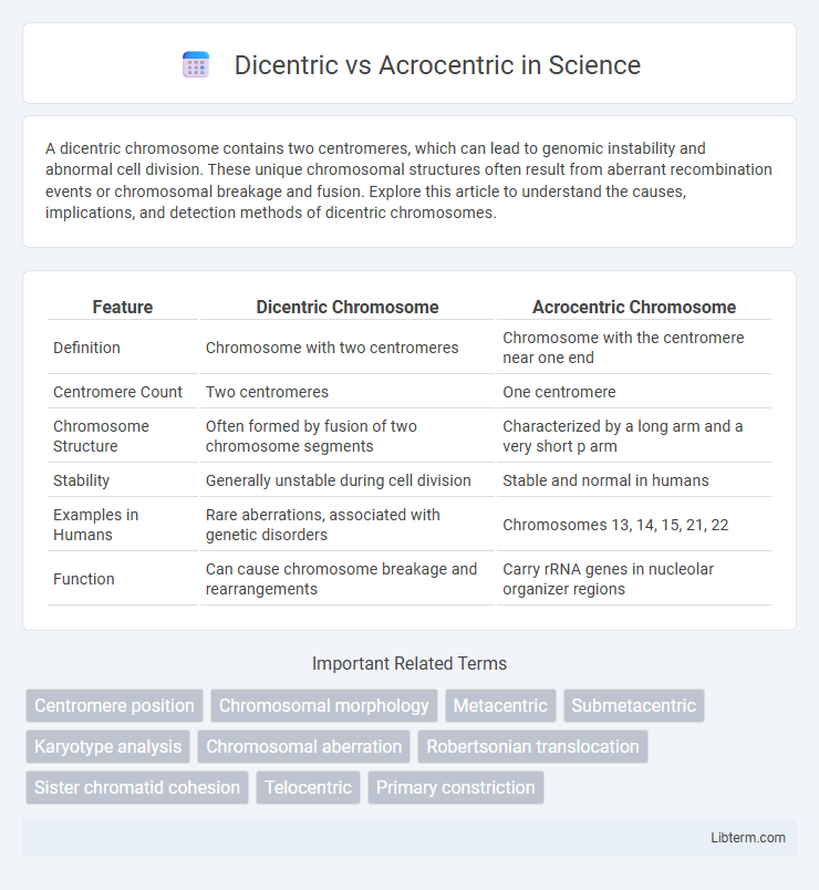
Table of Comparison
| Feature | Dicentric Chromosome | Acrocentric Chromosome |
|---|---|---|
| Definition | Chromosome with two centromeres | Chromosome with the centromere near one end |
| Centromere Count | Two centromeres | One centromere |
| Chromosome Structure | Often formed by fusion of two chromosome segments | Characterized by a long arm and a very short p arm |
| Stability | Generally unstable during cell division | Stable and normal in humans |
| Examples in Humans | Rare aberrations, associated with genetic disorders | Chromosomes 13, 14, 15, 21, 22 |
| Function | Can cause chromosome breakage and rearrangements | Carry rRNA genes in nucleolar organizer regions |
Introduction to Chromosome Structures
Dicentric chromosomes contain two centromeres, which can lead to instability during cell division due to the formation of chromosome bridges. Acrocentric chromosomes feature a single centromere positioned near one end, producing a long arm and a satellite-bearing short arm involved in ribosomal RNA gene clusters. Understanding the structural differences between dicentric and acrocentric chromosomes is crucial for studying chromosomal behavior, genetic stability, and their implications in genetic disorders.
Overview of Dicentric Chromosomes
Dicentric chromosomes contain two centromeres, which often result from abnormal chromosomal fusion events and can lead to chromosomal instability during cell division. These chromosomes are distinct from acrocentric chromosomes, which have a single centromere located near one end and typically feature short p arms with satellite DNA. The presence of two functional centromeres in dicentric chromosomes frequently causes anaphase bridges and chromosome breakage, contributing to genetic disorders and cancer development.
Understanding Acrocentric Chromosomes
Acrocentric chromosomes are characterized by having the centromere located very close to one end, resulting in a long arm and a satellite or short arm containing repetitive genetic material such as ribosomal RNA genes. This structure distinguishes them from dicentric chromosomes, which possess two centromeres and often lead to genomic instability due to abnormal chromosomal segregation. Understanding the unique morphology and genetic content of acrocentric chromosomes is essential for studying chromosomal behavior and related disorders, including Robertsonian translocations.
Key Structural Differences: Dicentric vs Acrocentric
Dicentric chromosomes possess two distinct centromeres, causing unique challenges during cell division, while acrocentric chromosomes feature a single centromere located near one end, producing a characteristic short p arm. Dicentric chromosomes often result from chromosomal fusion events, leading to instability or breakage, whereas acrocentric chromosomes typically carry nucleolar organizer regions essential for ribosomal RNA synthesis. The structural positioning of centromeres in dicentric chromosomes differs fundamentally from acrocentric chromosomes, impacting genetic behavior and chromosomal segregation.
Formation Mechanisms of Dicentric Chromosomes
Dicentric chromosomes form primarily through abnormal telomere end fusions or misrepair of DNA double-strand breaks, resulting in chromosomes with two centromeres. These events often involve chromosomal breakage followed by fusion at centromeric or subtelomeric regions, leading to chromosomal instability and potential anaphase bridges during cell division. In contrast, acrocentric chromosomes are structurally characterized by a centromere located near one end, involving different mechanisms related to centromere positioning rather than fusion events.
Biological Significance of Acrocentric Chromosomes
Acrocentric chromosomes hold key biological significance due to their unique structure featuring a centromere near one end, producing a very short p arm with genes crucial for ribosomal RNA production. These chromosomes participate prominently in nucleolus formation and contribute to maintaining genomic stability through processes like Robertsonian translocations, which can impact fertility and genetic disorders. Unlike dicentric chromosomes, which contain two centromeres and often lead to chromosomal instability, acrocentric chromosomes support essential cellular functions and genetic integrity through their specialized role.
Chromosomal Stability: Dicentric versus Acrocentric
Dicentric chromosomes contain two centromeres, often leading to chromosomal instability due to improper segregation during cell division, causing breakage and aneuploidy. Acrocentric chromosomes have a single centromere located near one end, which contributes to more stable inheritance patterns and reduced risk of breakage compared to dicentric chromosomes. The structural difference in centromere placement directly influences mitotic stability and genomic integrity in cellular processes.
Clinical Implications and Genetic Disorders
Dicentric chromosomes, characterized by two centromeres, often lead to genomic instability and are frequently associated with genetic disorders such as cancer and congenital abnormalities due to their propensity for chromosomal breakage and missegregation during cell division. Acrocentric chromosomes, which have a centromere near one end, are involved in Robertsonian translocations, a common form of chromosomal rearrangement linked to conditions like Down syndrome, Patau syndrome, and reproductive issues including infertility and miscarriage. Understanding the distinct clinical implications of dicentric and acrocentric chromosomes aids in diagnosing and managing chromosomal abnormalities and their related genetic disorders.
Diagnostic Techniques for Identifying Dicentric and Acrocentric Chromosomes
Fluorescence in situ hybridization (FISH) and G-banding karyotyping are primary diagnostic techniques for identifying dicentric and acrocentric chromosomes. Dicentric chromosomes are characterized by two centromeres detected using centromere-specific probes in FISH, while acrocentric chromosomes, identified by their distinctive short p arm and satellite DNA, are visualized in metaphase spreads with G-banding. Advanced methods like spectral karyotyping (SKY) enhance resolution, enabling precise differentiation and structural analysis of these chromosomal abnormalities.
Summary and Future Perspectives
Dicentric chromosomes contain two centromeres and often result in chromosomal instability, leading to cell division errors and genetic disorders, whereas acrocentric chromosomes have a centromere near one end and are characterized by short p arms with nucleolar organizer regions essential for ribosomal RNA production. Advances in molecular cytogenetics and live-cell imaging are enhancing the understanding of dicentric chromosome behavior during mitosis and their role in cancer development. Future research aims to elucidate the mechanisms of acrocentric chromosome rearrangements and their impact on genome organization and human disease susceptibility.
Dicentric Infographic