Immunohistochemistry utilizes antibodies to detect specific antigens in tissue samples, enabling precise localization of proteins and biomarkers. This technique plays a crucial role in diagnosing diseases, guiding treatment decisions, and advancing research by revealing cellular and molecular patterns. Explore the full article to understand how immunohistochemistry can enhance your diagnostic and research capabilities.
Table of Comparison
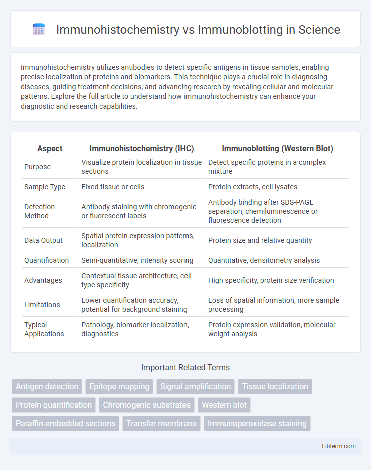
| Aspect | Immunohistochemistry (IHC) | Immunoblotting (Western Blot) |
|---|---|---|
| Purpose | Visualize protein localization in tissue sections | Detect specific proteins in a complex mixture |
| Sample Type | Fixed tissue or cells | Protein extracts, cell lysates |
| Detection Method | Antibody staining with chromogenic or fluorescent labels | Antibody binding after SDS-PAGE separation, chemiluminescence or fluorescence detection |
| Data Output | Spatial protein expression patterns, localization | Protein size and relative quantity |
| Quantification | Semi-quantitative, intensity scoring | Quantitative, densitometry analysis |
| Advantages | Contextual tissue architecture, cell-type specificity | High specificity, protein size verification |
| Limitations | Lower quantification accuracy, potential for background staining | Loss of spatial information, more sample processing |
| Typical Applications | Pathology, biomarker localization, diagnostics | Protein expression validation, molecular weight analysis |
Overview of Immunohistochemistry and Immunoblotting
Immunohistochemistry (IHC) utilizes antibodies to detect specific antigens in preserved tissue sections, enabling spatial localization and visualization of protein expression within the cellular context. Immunoblotting, commonly known as Western blotting, separates proteins by gel electrophoresis followed by antibody probing to quantify and identify target proteins in homogenized samples. Both techniques are critical in molecular biology for protein analysis, with IHC providing in situ information and immunoblotting offering precise molecular weight and expression level data.
Fundamental Principles of Each Technique
Immunohistochemistry relies on antigen-antibody interactions to visualize specific proteins within tissue sections using labeled antibodies and microscopy, preserving spatial context. Immunoblotting, or Western blotting, separates proteins by gel electrophoresis before transferring them to a membrane, where antibodies detect target proteins quantitatively. Both techniques utilize specific antigen recognition, but immunohistochemistry emphasizes localization while immunoblotting prioritizes protein identification and quantification.
Sample Preparation and Processing Differences
Immunohistochemistry (IHC) involves fixing tissue samples using formalin and embedding them in paraffin to preserve spatial morphology before sectioning for antibody staining, while immunoblotting (Western blotting) requires protein extraction from lysed cells or tissues followed by denaturation and separation via SDS-PAGE. IHC samples maintain structural context, enabling localization of target antigens within tissue architecture, whereas immunoblotting samples are homogenized, providing quantitative analysis of protein expression without cellular context. The fixation and embedding steps in IHC contrast sharply with the solubilization and electrophoretic transfer processes mandatory in immunoblotting workflows.
Detection Methods and Visualization
Immunohistochemistry (IHC) employs antibody-antigen interactions to detect specific proteins within fixed tissue sections, visualized through chromogenic or fluorescent labels that preserve spatial context. Immunoblotting, also known as Western blotting, detects proteins separated by gel electrophoresis using antibodies, with visualization achieved via chemiluminescent or colorimetric substrates generating signals on membranes. While IHC provides localization information within tissue architecture, immunoblotting offers quantitative and molecular weight data but lacks spatial resolution.
Sensitivity and Specificity Comparison
Immunohistochemistry (IHC) offers high specificity by localizing protein expression within intact tissue architecture, allowing precise cellular context identification, while immunoblotting (Western blot) provides enhanced sensitivity for detecting low-abundance proteins through protein separation and antibody probing. IHC's sensitivity can be affected by tissue fixation and antigen retrieval methods, whereas immunoblotting generally achieves greater sensitivity due to protein enrichment and isolation steps. Specificity in immunoblotting benefits from molecular weight confirmation, reducing false positives, whereas IHC specificity relies heavily on antibody quality and appropriate controls to minimize non-specific staining.
Applications in Biomedical Research
Immunohistochemistry enables precise localization of protein expression within tissue samples, making it essential for studying cellular context and tissue architecture in biomedical research. Immunoblotting, also known as Western blotting, quantifies protein levels and detects specific proteins in complex mixtures, facilitating analysis of protein expression, modification, and validation of antibodies. Both techniques complement each other by providing spatial and quantitative data crucial for understanding disease mechanisms and biomarker identification.
Advantages of Immunohistochemistry
Immunohistochemistry (IHC) offers precise localization of protein expression within the native tissue architecture, enabling visualization of spatial distribution and cellular context, which immunoblotting cannot provide. IHC is particularly advantageous for examining heterogeneous tissue samples and preserving morphological details while detecting specific antigens. This method facilitates in situ analysis, allowing correlation between protein presence and histopathological features, critical for diagnostic and research applications.
Strengths of Immunoblotting
Immunoblotting, also known as Western blotting, offers high specificity and sensitivity for detecting target proteins, enabling precise quantification and molecular weight determination. This technique excels in confirming protein identity and post-translational modifications through antibody binding, making it ideal for validating protein expression in complex samples. Immunoblotting's ability to separate proteins by electrophoresis prior to detection increases accuracy compared to Immunohistochemistry, which primarily provides spatial localization without detailed quantification.
Limitations and Challenges of Each Method
Immunohistochemistry (IHC) faces limitations such as tissue fixation artifacts that can mask antigen sites, leading to variable antibody binding and interpretation challenges due to subjective analysis. Immunoblotting, or Western blotting, struggles with issues like limited protein size resolution, potential cross-reactivity of antibodies, and inability to provide spatial localization within tissue samples. Both methods require rigorous antibody validation and controls to minimize false positives and ensure reproducible results.
Choosing the Right Technique for Your Research
Immunohistochemistry (IHC) enables localization of specific proteins within tissue sections, providing spatial context and cellular distribution, whereas immunoblotting (Western blot) quantifies protein expression levels in homogenized samples with high sensitivity. Selecting the appropriate technique depends on research goals: IHC is ideal for studying protein localization and tissue morphology, while immunoblotting suits precise quantification and detection of protein size or post-translational modifications. Researchers should consider sample type, required data resolution, and whether spatial information or protein quantification is paramount for experimental outcomes.
Immunohistochemistry Infographic