Ferroptotic cell death is a unique form of regulated cell demise characterized by iron-dependent lipid peroxidation, distinguishing it from apoptosis and necrosis. Understanding the molecular mechanisms driving ferroptosis can reveal novel therapeutic targets for diseases like cancer and neurodegeneration. Explore the rest of the article to learn how ferroptotic pathways influence your health and treatment options.
Table of Comparison
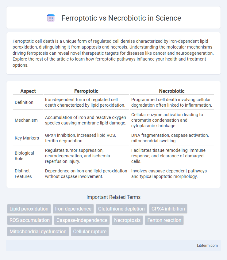
| Aspect | Ferroptotic | Necrobiotic |
|---|---|---|
| Definition | Iron-dependent form of regulated cell death characterized by lipid peroxidation. | Programmed cell death involving cellular degradation often linked to inflammation. |
| Mechanism | Accumulation of iron and reactive oxygen species causing membrane lipid damage. | Cellular enzyme activation leading to chromatin condensation and cytoplasmic shrinkage. |
| Key Markers | GPX4 inhibition, increased lipid ROS, ferritin degradation. | DNA fragmentation, caspase activation, mitochondrial swelling. |
| Biological Role | Regulates tumor suppression, neurodegeneration, and ischemia-reperfusion injury. | Facilitates tissue remodeling, immune response, and clearance of damaged cells. |
| Distinct Features | Dependence on iron and lipid peroxidation without caspase involvement. | Involves caspase-dependent pathways and typical apoptotic morphology. |
Introduction to Ferroptosis and Necrobiosis
Ferroptosis is a regulated form of cell death characterized by iron-dependent lipid peroxidation leading to membrane damage and cellular demise, distinct from traditional apoptosis and necrosis. Necrobiosis refers to the physiological or pathological degeneration and death of cells, often involving necrotic processes but lacking the specific biochemical markers of ferroptosis. Understanding the molecular pathways and triggers of ferroptosis and necrobiosis is critical for developing targeted therapies in diseases like cancer, neurodegeneration, and tissue injury.
Defining Ferroptotic Cell Death
Ferroptotic cell death is a regulated form of cell demise characterized by iron-dependent lipid peroxidation, distinguishing it from necrobiotic cell death, which involves cellular degeneration associated with inflammation and necrosis. Key molecular features of ferroptosis include glutathione depletion, inactivation of glutathione peroxidase 4 (GPX4), and accumulation of reactive oxygen species leading to membrane damage. This contrasts with necrobiosis, where cell death results from chronic injury or disease processes causing structural cellular breakdown without the iron-catalyzed lipid metabolic pathways central to ferroptosis.
Understanding Necrobiotic Processes
Necrobiotic processes involve the gradual breakdown and clearance of damaged or dying cells through mechanisms distinct from ferroptosis, which is characterized by iron-dependent lipid peroxidation leading to cell death. Necrobiosis primarily occurs in tissue remodeling and inflammatory responses, featuring cellular degradation without the extensive oxidative stress seen in ferroptosis. Understanding necrobiotic pathways enhances insights into chronic inflammation and degenerative diseases where controlled cell turnover is critical for maintaining tissue homeostasis.
Molecular Mechanisms: Ferroptosis vs Necrobiosis
Ferroptosis is a regulated cell death characterized by iron-dependent lipid peroxidation mediated by glutathione peroxidase 4 (GPX4) inactivation and accumulation of reactive oxygen species (ROS), triggering oxidative damage in cellular membranes. Necrobiosis involves programmed necrosis linked to ATP depletion and membrane rupture, often regulated by receptor-interacting protein kinases (RIPK1 and RIPK3) and mixed lineage kinase domain-like protein (MLKL) phosphorylation leading to plasma membrane disruption. While ferroptosis relies on iron metabolism and lipid peroxidation pathways, necrobiosis primarily depends on necroptotic signaling cascades involving kinase activity and controlled membrane lysis.
Key Biomarkers in Ferroptotic and Necrobiotic Pathways
Key biomarkers in ferroptotic pathways include elevated levels of lipid peroxides, glutathione depletion, and increased activity of glutathione peroxidase 4 (GPX4), all crucial for detecting iron-dependent cell death. Necrobiotic pathways are characterized by markers such as increased necrotic cell debris, elevated pro-inflammatory cytokines, and activation of receptor-interacting protein kinases (RIPK1 and RIPK3). Distinguishing these biomarkers allows for precise identification of ferroptotic versus necrobiotic cell death and contributes to targeted therapeutic strategies.
Cellular Morphology: Distinctions Between Ferroptosis and Necrobiosis
Ferroptosis is characterized by distinct cellular morphology including condensed mitochondrial membrane densities, reduced or vanished mitochondrial cristae, and outer mitochondrial membrane rupture, whereas necrobiosis demonstrates cellular swelling, plasma membrane rupture, and loss of nuclear integrity. In ferroptosis, lipid peroxidation leads to membrane damage without typical apoptotic features, contrasting with necrobiosis where enzymatic degradation causes cellular and tissue breakdown. These morphological differences are crucial for distinguishing ferroptotic death from necrobiotic processes in pathological examinations.
Pathophysiological Roles in Disease Contexts
Ferroptosis is an iron-dependent form of regulated cell death characterized by lipid peroxidation and accumulation of reactive oxygen species, playing a critical role in neurodegeneration, cancer, and ischemia-reperfusion injury. Necrobiotic cell death involves morphological changes such as cytoplasmic vacuolation and chromatin condensation, often associated with chronic inflammation and autoimmune diseases. Understanding the distinct molecular pathways of ferroptotic and necrobiotic processes aids in targeting pathological conditions like tumor resistance and inflammatory tissue damage.
Diagnostic Approaches: Differentiating Ferroptosis and Necrobiosis
Differentiating ferroptosis from necrobiosis relies on specific diagnostic approaches targeting distinct cellular and molecular markers. Ferroptosis is characterized by iron-dependent lipid peroxidation detectable via ferroptosis-specific probes such as C11-BODIPY and elevated levels of malondialdehyde, whereas necrobiosis shows hallmark morphological features like eosinophilic degeneration and is identified through histopathological staining techniques. Advanced methods including immunohistochemistry for glutathione peroxidase 4 (GPX4) depletion and transmission electron microscopy provide precise discrimination by revealing ferroptotic mitochondrial shrinkage versus necrobiotic cytoplasmic changes.
Therapeutic Implications and Intervention Strategies
Ferroptotic cell death, characterized by iron-dependent lipid peroxidation, offers therapeutic targets in diseases like cancer and neurodegeneration through modulation of iron metabolism and lipid antioxidants, while necrobiotic processes involving organized cell death and tissue remodeling suggest interventions targeting inflammatory pathways and extracellular matrix components. Ferroptosis inhibitors such as liproxstatin-1 and ferrostatin-1 show promise in limiting oxidative damage, whereas necrobiotic interventions prioritize anti-inflammatory agents and matrix metalloproteinase inhibitors to regulate tissue repair. Effective therapeutic strategies necessitate precise modulation of these distinct cell death mechanisms to control pathological progression and enhance tissue recovery.
Future Directions and Research Perspectives
Future directions in ferroptotic and necrobiotic research emphasize the development of targeted therapies exploiting distinct molecular pathways involved in lipid peroxidation and cellular membrane integrity. Advances in single-cell analysis and biomarker identification promise to enhance diagnostic precision and therapeutic monitoring for diseases linked to ferroptosis and necrobiosis. Investigating crosstalk between ferroptotic and necrobiotic mechanisms could unveil novel intervention points for complex pathologies such as neurodegeneration and cancer.
Ferroptotic Infographic