Necrosis is the uncontrolled death of cells or tissue caused by factors such as injury, infection, or lack of blood supply, leading to the breakdown of cell structures. This pathological process can result in inflammation and damage to surrounding healthy tissue, posing serious health risks if untreated. Discover the critical insights and treatments for necrosis to better understand how to protect your health in the full article.
Table of Comparison
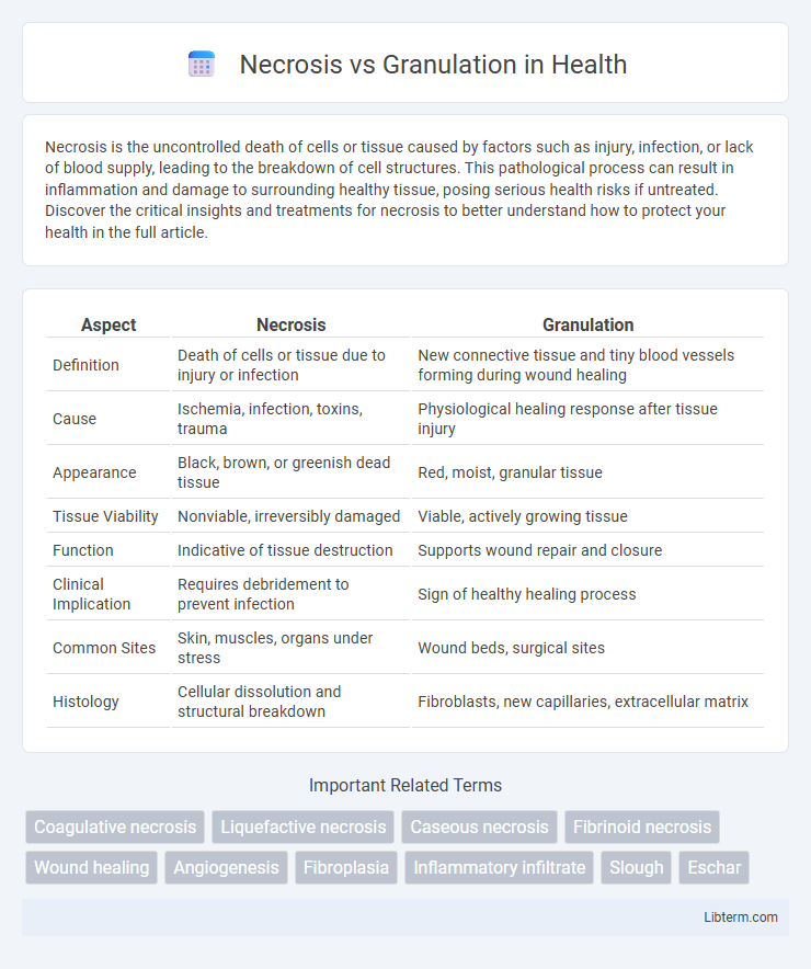
| Aspect | Necrosis | Granulation |
|---|---|---|
| Definition | Death of cells or tissue due to injury or infection | New connective tissue and tiny blood vessels forming during wound healing |
| Cause | Ischemia, infection, toxins, trauma | Physiological healing response after tissue injury |
| Appearance | Black, brown, or greenish dead tissue | Red, moist, granular tissue |
| Tissue Viability | Nonviable, irreversibly damaged | Viable, actively growing tissue |
| Function | Indicative of tissue destruction | Supports wound repair and closure |
| Clinical Implication | Requires debridement to prevent infection | Sign of healthy healing process |
| Common Sites | Skin, muscles, organs under stress | Wound beds, surgical sites |
| Histology | Cellular dissolution and structural breakdown | Fibroblasts, new capillaries, extracellular matrix |
Understanding Necrosis: Definition and Characteristics
Necrosis is the pathological death of cells or tissues due to factors like ischemia, infection, or toxins, characterized by loss of cell membrane integrity, swelling, and enzymatic digestion. It often results in inflammation and the release of cellular contents that can damage surrounding tissues. Understanding necrosis is critical for differentiating it from granulation tissue, which represents healthy repair with new capillary and fibroblast proliferation during wound healing.
What is Granulation Tissue? Key Features Explained
Granulation tissue is newly formed connective tissue and microscopic blood vessels that develop on the surfaces of a wound during the healing process. It is characterized by a rich extracellular matrix, proliferating fibroblasts, and abundant capillaries, which facilitate oxygen and nutrient delivery essential for tissue repair. Unlike necrosis, which involves cellular death and tissue destruction, granulation tissue signifies healthy wound healing and the body's response to injury.
Causes of Necrosis in Wound Healing
Necrosis in wound healing primarily results from factors such as prolonged ischemia, infection, and severe trauma that disrupt blood supply and cellular integrity. Cellular death occurs due to oxygen deprivation, accumulation of toxic metabolites, and inflammatory responses that overwhelm tissue repair mechanisms. Understanding these causes helps differentiate necrotic tissue from granulation tissue, the latter characterized by new capillary formation and fibroblast proliferation essential for wound closure.
The Role of Granulation in Tissue Repair
Granulation tissue plays a crucial role in tissue repair by providing a vascularized matrix that supports cell proliferation and wound healing, contrasting with necrosis where dead tissue impedes recovery. This newly formed connective tissue contains fibroblasts, immune cells, and proliferating capillaries essential for replacing damaged cells and restoring tissue integrity. Effective granulation prevents infection and promotes re-epithelialization, making it a vital phase in the regenerative process following injury.
Visual Differences: Necrosis vs Granulation
Necrosis appears as black, brown, or dark green tissue indicating dead cells, often dry or moist with a firm or soft texture, while granulation tissue is characterized by bright red or pink, moist, and bumpy appearance due to new capillary growth. Necrotic tissue lacks vascularization and is typically surrounded by inflammation or infection, whereas granulation tissue is well-vascularized, indicating healing and tissue regeneration. Visual examination distinguishes necrosis by its discolored, devitalized areas contrasted with granulation's healthy, vibrant red matrix essential for wound repair.
Cellular and Molecular Mechanisms
Necrosis involves uncontrolled cell death characterized by membrane rupture, release of intracellular contents, and induction of inflammation through the activation of damage-associated molecular patterns (DAMPs) like HMGB1. Granulation tissue formation occurs through the coordinated proliferation of fibroblasts, endothelial cells, and inflammatory cells, driven by growth factors such as VEGF, TGF-b, and PDGF, promoting angiogenesis and extracellular matrix deposition. Cellular signaling pathways including NF-kB and MAPK regulate the inflammatory response in necrosis, while granulation tissue relies on cytokines and chemokines to mediate tissue repair and remodeling.
Clinical Implications of Necrosis and Granulation
Necrosis indicates irreversible cell death leading to tissue destruction, often resulting in infection, delayed healing, and increased morbidity in clinical settings. Granulation tissue formation represents a critical phase of wound healing characterized by new capillaries, fibroblasts, and extracellular matrix, promoting tissue repair and restoration of function. Recognizing necrotic tissue requires prompt debridement to prevent chronic wounds, while granulation signals effective healing progression and guides therapeutic interventions.
Diagnosis: How to Differentiate Necrotic and Granulation Tissue
Differentiating necrotic from granulation tissue relies heavily on visual assessment and diagnostic tools. Necrotic tissue appears black or brown, is non-viable, and often has a dry, leathery texture, while granulation tissue is red or pink, moist, and composed of new connective tissue and capillaries indicating healing. Advanced techniques such as wound biopsy, histopathology, and imaging modalities like infrared thermography can enhance accuracy in distinguishing these tissues for appropriate wound management.
Treatment Strategies for Necrosis and Granulation Management
Treatment strategies for necrosis focus on timely debridement to remove dead tissue, preventing infection and promoting wound healing. Granulation management emphasizes maintaining a moist wound environment, supporting angiogenesis, and using topical agents like growth factors or antimicrobial dressings to enhance tissue regeneration. Both conditions require careful assessment to optimize healing outcomes and minimize complications.
Prognosis and Outcomes: Impact on Wound Healing
Necrosis impedes wound healing by creating a barrier to re-epithelialization and increasing the risk of infection, often resulting in delayed recovery or chronic wounds. Granulation tissue, composed of new connective tissue and microscopic blood vessels, promotes wound closure and signifies a positive healing trajectory with improved prognosis. Effective debridement of necrotic tissue enhances the development of granulation and overall wound outcomes.
Necrosis Infographic