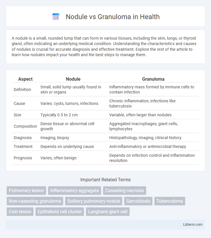
A nodule is a small, rounded lump that can form in various tissues, including the skin, lungs, or thyroid gland, often indicating an underlying medical condition. Understanding the characteristics and causes of nodules is crucial for accurate diagnosis and effective treatment. Explore the rest of the article to learn how nodules impact your health and the best steps to manage them.
Table of Comparison
| Aspect | Nodule | Granuloma |
|---|---|---|
| Definition | Small, solid lump usually found in skin or organs | Inflammatory mass formed by immune cells to contain infection |
| Cause | Varies: cysts, tumors, infections | Chronic inflammation, infections like tuberculosis |
| Size | Typically 0.5 to 2 cm | Variable, often larger than nodules |
| Composition | Dense tissue or abnormal cell growth | Aggregated macrophages, giant cells, lymphocytes |
| Diagnosis | Imaging, biopsy | Histopathology, imaging, clinical history |
| Treatment | Depends on underlying cause | Anti-inflammatory or antimicrobial therapy |
| Prognosis | Varies, often benign | Depends on infection control and inflammation resolution |
Introduction to Nodules and Granulomas
Nodules are small, rounded masses of tissue that can develop in various organs due to inflammation, infection, or neoplastic processes. Granulomas represent a specific type of nodular inflammation characterized by a focused collection of macrophages, often forming in response to persistent infections like tuberculosis or chronic inflammatory conditions. Differentiating between nodules and granulomas is critical for accurate diagnosis and treatment, as granulomas indicate an immune-mediated response while nodules encompass a broader range of etiologies.
Defining Nodules: Key Characteristics
Nodules are small, firm, rounded masses typically measuring between 1 cm and 3 cm in diameter, often found within the skin, lungs, or thyroid gland. They differ from granulomas in that nodules represent a broader category of lesions that can be benign, malignant, or inflammatory, whereas granulomas specifically consist of organized collections of macrophages formed in response to chronic inflammation or infection. Key characteristics of nodules include their solid consistency, well-defined borders, and potential to cause localized tissue displacement or dysfunction.
Understanding Granulomas: An Overview
Granulomas are organized collections of immune cells, primarily macrophages, that form as a response to chronic inflammation, infection, or foreign substances, distinct from nodules which are solid lesions often detected in imaging studies. Granulomas serve as a protective mechanism, isolating harmful agents to prevent their spread, with common causes including tuberculosis, sarcoidosis, and certain fungal infections. Histologically, granulomas are characterized by epithelioid cells, multinucleated giant cells, and a surrounding lymphocytic cuff, differentiating them from simpler nodular structures.
Causes of Nodules vs Granulomas
Nodules often arise from benign or malignant neoplasms, infectious processes like tuberculosis, or inflammatory conditions such as rheumatoid arthritis. Granulomas form primarily due to chronic inflammatory responses triggered by persistent infections, including mycobacterial bacteria, fungal organisms, or foreign substances like silica and beryllium. The key difference lies in nodules representing a broad category of solid lesions, while granulomas specifically indicate organized collections of macrophages responding to difficult-to-eradicate antigens.
Clinical Presentation and Symptoms
Nodules typically present as palpable, firm lumps beneath the skin or within organs, often asymptomatic but sometimes causing pain or discomfort depending on size and location. Granulomas manifest as localized inflammatory lesions characterized by immune cell aggregation, frequently associated with chronic infections or autoimmune conditions, leading to symptoms such as persistent cough, fever, or weight loss when occurring in the lungs. Clinical presentation varies significantly, with nodules often detected incidentally during imaging, whereas granulomas may present with systemic symptoms reflecting underlying disease processes.
Diagnostic Approaches: Nodules vs Granulomas
Diagnostic approaches for nodules involve imaging techniques such as ultrasound, CT scans, and MRI to assess size, shape, and location, often followed by fine-needle aspiration or biopsy for histopathological evaluation. Granulomas require tissue biopsy to identify characteristic clusters of macrophages, often supported by special staining or molecular tests to detect infectious agents like Mycobacterium tuberculosis or fungal infections. Differential diagnosis relies heavily on imaging combined with histological analysis to distinguish benign nodules from granulomatous inflammation linked to chronic infections or autoimmune conditions.
Imaging Findings and Differences
Nodules on imaging often appear as well-defined, round or oval lesions with uniform density, commonly seen on chest X-rays or CT scans, whereas granulomas typically present as small, dense, calcified nodules resulting from chronic inflammation or infection. Granulomas usually have a characteristic pattern of central necrosis or calcification visible on imaging, distinguishing them from benign or malignant nodules that lack such features. Imaging modalities like CT scans provide detailed contrast differences, aiding in differentiating granulomas caused by diseases like tuberculosis or sarcoidosis from neoplastic nodules.
Histopathological Features
Nodules are localized collections of cells typically characterized by dense fibrous tissue and an accumulation of inflammatory cells, while granulomas present as organized aggregates of macrophages, often transforming into epithelioid cells, surrounded by lymphocytes and occasional multinucleated giant cells. Histopathologically, granulomas exhibit a distinct architecture with caseating or non-caseating necrosis, depending on the underlying etiology, whereas nodules lack this structured immune response and necrosis pattern. Identification of cellular composition and necrosis type through biopsy and staining techniques is critical in differentiating nodules from granulomas in clinical diagnosis.
Treatment Options for Nodules and Granulomas
Treatment options for nodules typically include corticosteroid injections, physical therapy, or surgical excision if the nodule causes significant symptoms or functional impairment. Granulomas often require addressing the underlying cause, such as infection or inflammation, with antibiotics, antifungal medications, or corticosteroids to reduce immune response. In refractory cases, surgical removal or immunosuppressive therapies may be necessary to resolve persistent granulomatous inflammation.
Prognosis and Potential Complications
Nodules typically have a favorable prognosis when benign, often requiring minimal intervention unless they impact surrounding structures, while granulomas may indicate chronic inflammation or infection, potentially leading to fibrosis or tissue damage if untreated. Granulomas, particularly those caused by persistent infections like tuberculosis or sarcoidosis, can result in complications such as organ dysfunction or chronic granulomatous disease. Early diagnosis and management improve outcomes by preventing progression to irreversible tissue injury or systemic involvement.
Nodule Infographic