Atrophy refers to the gradual decline or wasting away of tissues, muscles, or organs due to disease, injury, or lack of use. It often results in reduced strength, mobility, or function, severely impacting overall health and quality of life. Discover how atrophy develops, its causes, symptoms, and effective treatments by reading the full article.
Table of Comparison
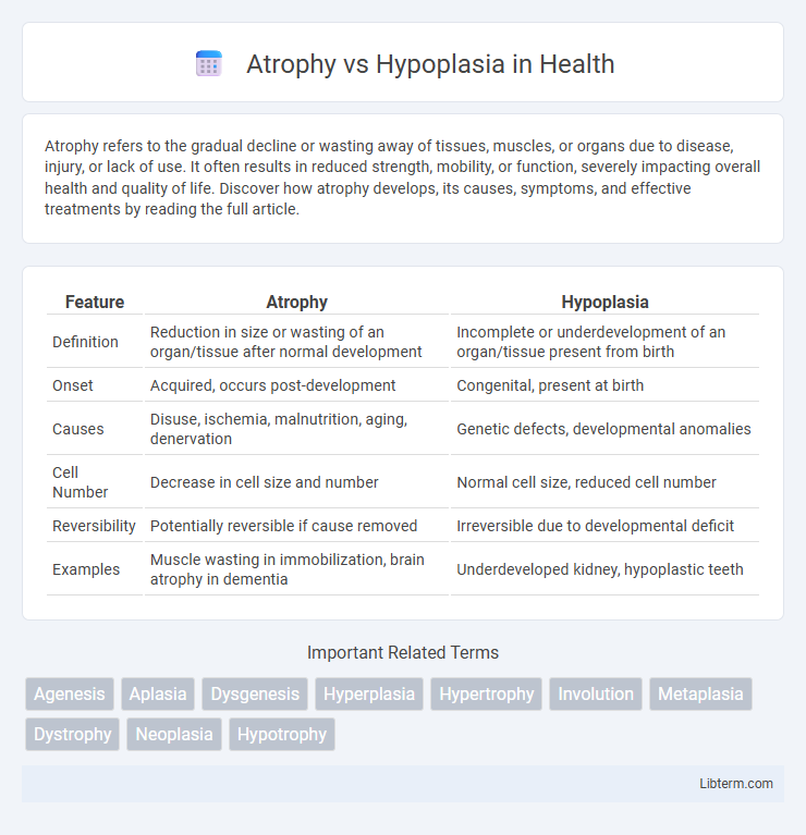
| Feature | Atrophy | Hypoplasia |
|---|---|---|
| Definition | Reduction in size or wasting of an organ/tissue after normal development | Incomplete or underdevelopment of an organ/tissue present from birth |
| Onset | Acquired, occurs post-development | Congenital, present at birth |
| Causes | Disuse, ischemia, malnutrition, aging, denervation | Genetic defects, developmental anomalies |
| Cell Number | Decrease in cell size and number | Normal cell size, reduced cell number |
| Reversibility | Potentially reversible if cause removed | Irreversible due to developmental deficit |
| Examples | Muscle wasting in immobilization, brain atrophy in dementia | Underdeveloped kidney, hypoplastic teeth |
Understanding Atrophy: Definition and Causes
Atrophy refers to the decrease in size or wasting away of an organ or tissue due to the reduction in the size or number of its cells, commonly caused by factors such as aging, malnutrition, ischemia, or disuse. Unlike hypoplasia, which is a congenital underdevelopment or incomplete development of a tissue or organ, atrophy is an acquired condition occurring after normal development. Understanding atrophy involves recognizing its reversible nature in early stages and the role of cell degradation pathways such as autophagy and apoptosis in its progression.
What is Hypoplasia? Key Features and Origins
Hypoplasia is a developmental condition characterized by the underdevelopment or incomplete formation of an organ or tissue due to a reduced number of cells. Key features of hypoplasia include smaller size, lack of normal function, and structural immaturity, often evident from birth. Origins of hypoplasia typically involve genetic mutations, prenatal exposure to toxins, or disrupted blood supply during critical periods of fetal development.
Atrophy vs Hypoplasia: Core Differences
Atrophy involves the reduction in size or wasting away of existing cells or tissues due to factors like aging, malnutrition, or disuse, resulting in decreased organ or tissue volume. Hypoplasia refers to the underdevelopment or incomplete formation of an organ or tissue during embryonic growth, characterized by fewer cells or structural deficiencies from birth. The core difference lies in atrophy being a post-developmental degeneration process, whereas hypoplasia is a congenital defect caused by impaired developmental processes.
Common Conditions Associated with Atrophy
Atrophy commonly occurs in conditions such as muscle disuse, aging (sarcopenia), malnutrition, and neurodegenerative diseases like Alzheimer's and Parkinson's. It is also frequently seen in ischemic tissues, endocrine disorders (e.g., adrenal atrophy), and chronic inflammatory states leading to tissue shrinkage. Hypoplasia, in contrast, is a developmental condition characterized by incomplete tissue or organ formation rather than degeneration.
Hypoplasia in Developmental Disorders
Hypoplasia refers to the underdevelopment or incomplete formation of an organ or tissue during fetal development, often leading to smaller size and reduced functionality compared to normal growth standards. In developmental disorders, hypoplasia is a critical factor in conditions such as hypoplastic left heart syndrome, where the left side of the heart is underdeveloped, causing severe cardiovascular complications. Unlike atrophy, which involves the degeneration of previously developed tissue, hypoplasia is characterized by an intrinsic failure of growth, impacting organs like kidneys, brain, or teeth during embryogenesis.
Clinical Signs: Identifying Atrophy and Hypoplasia
Atrophy presents clinically as a reduction in the size and function of previously normal tissue or organ, often accompanied by muscle weakness, decreased mass, and skin thinning in affected areas. Hypoplasia is identified by the underdevelopment or incomplete formation of an organ or tissue, detectable at birth or early childhood, with signs varying based on the specific tissue involved, such as hypoplastic enamel in teeth or hypoplastic muscles causing functional deficits. Distinguishing between atrophy and hypoplasia relies on patient history, physical examination, and imaging studies showing tissue regression in atrophy versus congenital size reduction in hypoplasia.
Diagnostic Approaches for Atrophy and Hypoplasia
Diagnostic approaches for atrophy primarily involve imaging techniques such as MRI and CT scans to detect tissue shrinkage, alongside biopsy for histopathological confirmation. In hypoplasia, diagnosis relies on imaging studies to identify underdeveloped organs or tissues, often supplemented by genetic testing to determine congenital causes. Both conditions require detailed clinical evaluation and comparison with normal anatomical development to differentiate functional deficits from structural abnormalities.
Treatment Strategies for Atrophy
Treatment strategies for atrophy primarily involve addressing the underlying cause, such as improving nutrition, enhancing blood flow, or reducing nerve damage. Physical therapy and exercise are often recommended to stimulate muscle growth and prevent further tissue loss. In some cases, pharmacological interventions like anti-inflammatory drugs or hormone replacement therapy may be employed to support tissue regeneration and functional recovery.
Managing Hypoplasia: Therapeutic Options
Managing hypoplasia involves targeted therapeutic options such as growth factor therapy, stem cell treatment, and surgical interventions to stimulate tissue development and enhance functional capacity. Early diagnosis allows for personalized treatment plans incorporating physical therapy and nutritional support to maximize growth potential. Emerging regenerative medicine techniques aim to restore hypoplastic tissues by promoting cellular proliferation and differentiation.
Prognosis and Long-Term Outcomes
Atrophy involves the reduction in size of previously normal tissue due to cell loss or shrinkage, often leading to variable but potentially reversible functional impairment depending on the cause and duration. Hypoplasia refers to the incomplete development or underdevelopment of a tissue or organ, resulting in permanently reduced cell numbers and typically irreversible functional deficits. Prognosis for atrophy generally improves with early intervention and removal of offending factors, whereas hypoplasia often results in lifelong limitations due to structural deficits established during development.
Atrophy Infographic