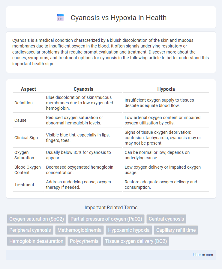
Cyanosis is a medical condition characterized by a bluish discoloration of the skin and mucous membranes due to insufficient oxygen in the blood. It often signals underlying respiratory or cardiovascular problems that require prompt evaluation and treatment. Discover more about the causes, symptoms, and treatment options for cyanosis in the following article to better understand this important health sign.
Table of Comparison
| Aspect | Cyanosis | Hypoxia |
|---|---|---|
| Definition | Blue discoloration of skin/mucous membranes due to low oxygenated hemoglobin. | Insufficient oxygen supply to tissues despite adequate blood flow. |
| Cause | Reduced oxygen saturation or abnormal hemoglobin levels. | Low arterial oxygen content or impaired oxygen utilization by cells. |
| Clinical Sign | Visible blue tint, especially in lips, fingers, toes. | Signs of tissue oxygen deprivation: confusion, tachycardia, cyanosis may or may not be present. |
| Oxygen Saturation | Usually below 85% for cyanosis to appear. | Can be normal or low; depends on underlying cause. |
| Blood Oxygen Content | Decreased oxygenated hemoglobin concentration. | Low oxygen delivery or impaired oxygen usage. |
| Treatment | Address underlying cause, oxygen therapy if needed. | Restore adequate oxygen delivery and consumption. |
Definition of Cyanosis
Cyanosis is a bluish discoloration of the skin and mucous membranes caused by an increased concentration of deoxygenated hemoglobin in the blood, typically when it exceeds 5 g/dL. It is a clinical sign indicating underlying hypoxemia but does not directly measure oxygen deficiency at the tissue level. Unlike hypoxia, which refers to inadequate oxygen supply to tissues, cyanosis specifically reflects the visible changes due to reduced oxygen saturation in the blood.
Definition of Hypoxia
Hypoxia is a medical condition characterized by an insufficient supply of oxygen to the tissues, despite adequate blood oxygen levels. It differs from cyanosis, which is the bluish discoloration of the skin and mucous membranes caused by increased deoxygenated hemoglobin in the blood. Hypoxia can result from various causes, including respiratory failure, anemia, or circulatory problems, and requires prompt diagnosis and treatment to prevent tissue damage.
Causes of Cyanosis
Cyanosis results from an increased amount of deoxygenated hemoglobin in the blood, often caused by conditions like chronic obstructive pulmonary disease (COPD), congenital heart defects, or severe pulmonary embolism. Hypoxia refers to a deficiency in oxygen reaching tissues, which can stem from anemia, carbon monoxide poisoning, or high altitude exposure. Unlike hypoxia, cyanosis specifically indicates visible bluish discoloration due to impaired oxygenation in peripheral tissues.
Causes of Hypoxia
Hypoxia results from inadequate oxygen supply to tissues due to causes such as respiratory disorders (e.g., chronic obstructive pulmonary disease, asthma), cardiovascular conditions (e.g., heart failure, shock), anemia, or high altitude exposure. It can also arise from impaired oxygen utilization at the cellular level, including mitochondrial dysfunction or poisoning by substances like cyanide. Differentiating hypoxia from cyanosis is crucial, as hypoxia reflects oxygen deprivation at the tissue level, while cyanosis indicates the bluish discoloration of skin caused by increased deoxygenated hemoglobin in the blood.
Pathophysiological Differences
Cyanosis results from increased levels of deoxygenated hemoglobin (above 5 g/dL) in the capillary blood, causing a bluish discoloration of the skin and mucous membranes, while hypoxia refers to reduced oxygen availability at the tissue level regardless of oxygen content in the blood. Pathophysiologically, cyanosis is primarily a visible sign of hypoxemia or abnormal hemoglobin function, whereas hypoxia involves impaired oxygen delivery due to factors such as low arterial oxygen tension, anemia, or circulatory defects. Understanding these differences is critical for accurate diagnosis and treatment, focusing on either improving blood oxygenation or enhancing tissue oxygen utilization.
Clinical Signs and Symptoms
Cyanosis presents as a bluish discoloration of the skin and mucous membranes, primarily indicating decreased oxygen saturation in capillary blood, commonly seen in peripheral tissues like lips, fingers, and toes. Hypoxia refers to inadequate oxygen supply to tissues and can manifest with symptoms such as shortness of breath, tachycardia, confusion, and cyanosis, though cyanosis may be absent in mild or early hypoxia. Clinical differentiation involves assessing oxygen saturation levels, respiratory rate, and neurological signs to identify underlying causes and severity.
Diagnostic Approaches
Cyanosis is identified through physical examination, revealing bluish discoloration of the skin or mucous membranes due to increased deoxygenated hemoglobin, whereas hypoxia requires blood gas analysis to measure arterial oxygen tension (PaO2) and oxygen saturation. Pulse oximetry provides a non-invasive method to estimate oxygen saturation but may not distinguish between cyanosis and hypoxia effectively. Advanced diagnostic tools such as arterial blood gas (ABG) analysis and co-oximetry are essential for accurate differentiation, guiding appropriate clinical management.
Treatment Strategies
Treatment strategies for cyanosis focus on improving oxygen delivery to tissues by administering supplemental oxygen and addressing underlying causes such as cardiac or pulmonary disorders. Hypoxia management involves identifying and correcting the root cause, including optimizing ventilation, treating anemia, or improving cardiac output to enhance systemic oxygenation. Both conditions require prompt intervention to prevent organ damage, often using targeted therapies like mechanical ventilation, medications, or surgical correction depending on the etiology.
Prognosis and Complications
Cyanosis indicates reduced oxygen saturation in hemoglobin but does not always correlate with tissue hypoxia, whereas hypoxia represents inadequate oxygen delivery to tissues, leading to cellular dysfunction and organ damage. Prognosis in cyanosis depends on the underlying cause and severity, with persistent hypoxia increasing risks of complications such as heart failure, neurological deficits, and metabolic acidosis. Early detection and management of hypoxia are critical to prevent irreversible tissue injury and improve clinical outcomes.
Key Differences Between Cyanosis and Hypoxia
Cyanosis is a physical sign characterized by a bluish discoloration of the skin and mucous membranes due to an increased concentration of deoxygenated hemoglobin in blood vessels near the surface. Hypoxia refers to a condition where tissues are deprived of adequate oxygen supply, impacting cellular metabolism and function regardless of blood color. While cyanosis indicates a visible change linked to oxygen saturation levels below 85%, hypoxia encompasses a broader physiological state that may not always present with skin discoloration.
Cyanosis Infographic

 libterm.com