Hypochromia is a condition characterized by red blood cells that have less color than normal, indicating lower hemoglobin levels. This often points to underlying issues such as iron deficiency anemia or other forms of anemia that affect oxygen transport in the body. Discover how hypochromia impacts your health and the best ways to address it by reading the rest of the article.
Table of Comparison
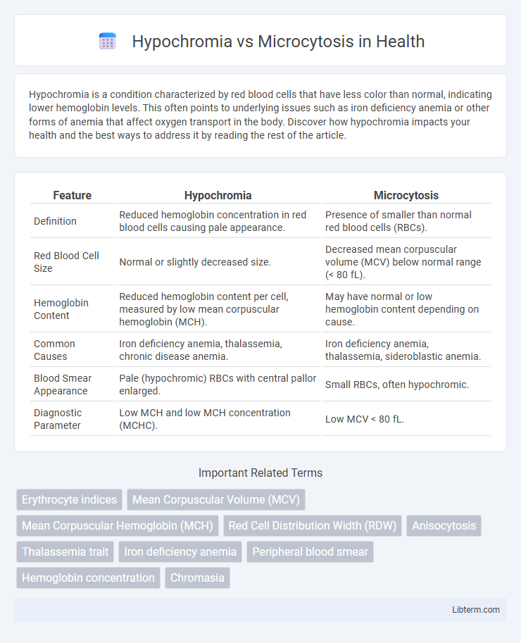
| Feature | Hypochromia | Microcytosis |
|---|---|---|
| Definition | Reduced hemoglobin concentration in red blood cells causing pale appearance. | Presence of smaller than normal red blood cells (RBCs). |
| Red Blood Cell Size | Normal or slightly decreased size. | Decreased mean corpuscular volume (MCV) below normal range (< 80 fL). |
| Hemoglobin Content | Reduced hemoglobin content per cell, measured by low mean corpuscular hemoglobin (MCH). | May have normal or low hemoglobin content depending on cause. |
| Common Causes | Iron deficiency anemia, thalassemia, chronic disease anemia. | Iron deficiency anemia, thalassemia, sideroblastic anemia. |
| Blood Smear Appearance | Pale (hypochromic) RBCs with central pallor enlarged. | Small RBCs, often hypochromic. |
| Diagnostic Parameter | Low MCH and low MCH concentration (MCHC). | Low MCV < 80 fL. |
Introduction to Hypochromia and Microcytosis
Hypochromia refers to red blood cells displaying reduced hemoglobin content, resulting in paler cells visible on a blood smear, commonly associated with iron deficiency anemia. Microcytosis describes the presence of smaller-than-normal red blood cells, often identified by a decreased mean corpuscular volume (MCV), and is typical in conditions like thalassemia and iron deficiency. Both hypochromia and microcytosis are key hematological indicators used to diagnose and differentiate types of anemia based on cell size and color intensity.
Defining Hypochromia: Causes and Significance
Hypochromia is characterized by red blood cells with reduced hemoglobin concentration, often identified by pale staining on blood smears, primarily caused by iron deficiency anemia, thalassemia, or chronic diseases. Its significance lies in indicating impaired hemoglobin synthesis, reflecting inadequate oxygen delivery capacity and guiding diagnostic evaluation for underlying conditions. Differentiating hypochromia from microcytosis, which involves smaller than normal red blood cells, helps pinpoint specific hematologic disorders requiring targeted treatment.
Understanding Microcytosis: Key Characteristics
Microcytosis is characterized by the presence of smaller than normal red blood cells, often indicated by a reduced mean corpuscular volume (MCV) below 80 fL. It is commonly associated with conditions such as iron deficiency anemia, thalassemia, and chronic disease anemia, where impaired hemoglobin synthesis leads to decreased cell size. Differentiating microcytosis from hypochromia involves analyzing cell size versus hemoglobin concentration, as hypochromia specifically refers to reduced hemoglobin content resulting in pale red blood cells.
Pathophysiological Mechanisms: Hypochromia vs Microcytosis
Hypochromia results from decreased hemoglobin concentration within red blood cells, often linked to impaired hemoglobin synthesis seen in iron deficiency anemia or thalassemia. Microcytosis involves a reduction in red blood cell size caused by defective hemoglobin production or disrupted erythropoiesis affecting cell maturation. Both conditions reflect underlying disruptions in hemoglobin production pathways, but hypochromia primarily impacts hemoglobin content, whereas microcytosis affects red cell morphology.
Common Disorders Associated with Hypochromia
Hypochromia is commonly associated with iron deficiency anemia, thalassemia, and sideroblastic anemia, characterized by reduced hemoglobin content in red blood cells. Unlike microcytosis, which involves smaller than normal red blood cells, hypochromia specifically indicates decreased pigmentation due to insufficient hemoglobin synthesis. Disorders such as chronic diseases, lead poisoning, and certain genetic conditions also frequently present with hypochromic red blood cells.
Conditions Frequently Linked to Microcytosis
Microcytosis commonly occurs in conditions such as iron deficiency anemia, thalassemia, and anemia of chronic disease, where red blood cells are smaller than normal due to impaired hemoglobin synthesis. Hypochromia, characterized by reduced hemoglobin concentration within red blood cells, often accompanies microcytosis but can also occur independently in iron deficiency states. Differentiating these conditions through parameters like mean corpuscular volume (MCV) and mean corpuscular hemoglobin concentration (MCHC) is crucial for accurate diagnosis and targeted treatment.
Diagnostic Evaluation: Laboratory Findings
Hypochromia is characterized by reduced hemoglobin concentration in red blood cells, often identified through decreased mean corpuscular hemoglobin (MCH) levels on a complete blood count (CBC). Microcytosis presents as smaller than normal red blood cells, reflected in a decreased mean corpuscular volume (MCV) on laboratory evaluation. Both conditions commonly appear in iron deficiency anemia and thalassemia, requiring serum ferritin, iron studies, and hemoglobin electrophoresis to differentiate underlying causes accurately.
Differential Diagnosis: Distinguishing Features
Hypochromia and microcytosis are key hematological features often evaluated in the differential diagnosis of anemia, with hypochromia indicating reduced hemoglobin concentration in red blood cells and microcytosis referring to smaller than normal red blood cell size. Hypochromia is predominantly associated with iron deficiency anemia and thalassemia traits, whereas microcytosis can also be seen in chronic diseases and lead poisoning, making red blood cell indices like MCV and MCH crucial for differentiation. Distinguishing features include normal or low serum ferritin levels in iron deficiency anemia causing hypochromia and elevated HbA2 in beta-thalassemia traits leading to microcytosis, guiding targeted diagnostic and treatment approaches.
Clinical Implications and Treatment Approaches
Hypochromia, characterized by decreased hemoglobin concentration in red blood cells, often indicates iron deficiency anemia, while microcytosis refers to smaller than normal red blood cells, commonly seen in thalassemia and iron deficiency anemia. Differentiating between hypochromia and microcytosis through blood smear analysis and iron studies is essential for accurate diagnosis and targeted treatment strategies. Treatment approaches vary, with iron supplementation being primary for hypochromic iron deficiency anemia, whereas microcytosis due to genetic conditions may require more specialized management including genetic counseling and transfusion therapy.
Summary: Key Takeaways and Future Perspectives
Hypochromia is characterized by reduced hemoglobin concentration in red blood cells, leading to pale cell appearance, while microcytosis involves smaller-than-normal red blood cells, often linked to conditions like iron deficiency anemia and thalassemia. Key takeaways highlight their diagnostic importance in differentiating hematologic disorders, with hypochromia impacting oxygen transport efficiency and microcytosis indicating altered erythropoiesis. Future perspectives emphasize advanced molecular diagnostics and personalized treatment strategies to improve patient outcomes through precise characterization of these erythrocyte abnormalities.
Hypochromia Infographic