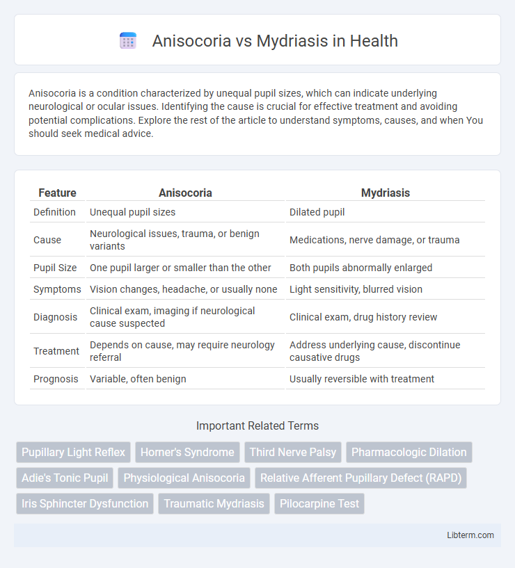
Anisocoria is a condition characterized by unequal pupil sizes, which can indicate underlying neurological or ocular issues. Identifying the cause is crucial for effective treatment and avoiding potential complications. Explore the rest of the article to understand symptoms, causes, and when You should seek medical advice.
Table of Comparison
| Feature | Anisocoria | Mydriasis |
|---|---|---|
| Definition | Unequal pupil sizes | Dilated pupil |
| Cause | Neurological issues, trauma, or benign variants | Medications, nerve damage, or trauma |
| Pupil Size | One pupil larger or smaller than the other | Both pupils abnormally enlarged |
| Symptoms | Vision changes, headache, or usually none | Light sensitivity, blurred vision |
| Diagnosis | Clinical exam, imaging if neurological cause suspected | Clinical exam, drug history review |
| Treatment | Depends on cause, may require neurology referral | Address underlying cause, discontinue causative drugs |
| Prognosis | Variable, often benign | Usually reversible with treatment |
Understanding Pupillary Abnormalities
Anisocoria refers to a condition where the pupils are uneven in size, which may indicate underlying neurological issues or eye trauma. Mydriasis is characterized by an abnormally dilated pupil that persists despite changes in lighting, often caused by drug effects, nerve damage, or brain injury. Differentiating between anisocoria and mydriasis is crucial for diagnosing pupillary abnormalities and assessing the integrity of the autonomic nervous system.
Defining Anisocoria: Causes and Significance
Anisocoria is a condition characterized by an unequal pupil size between the two eyes, which can result from physiological variations or pathological causes such as Horner's syndrome, Adie's tonic pupil, or third nerve palsy. Distinguishing anisocoria from mydriasis, which specifically refers to an abnormally dilated pupil, is crucial for accurate diagnosis and treatment. Recognition of anisocoria's causes and clinical significance helps identify underlying neurological or ocular disorders requiring prompt medical attention.
What is Mydriasis? Common Triggers and Mechanisms
Mydriasis is the abnormal dilation of the pupil caused by the relaxation of the iris sphincter muscle or stimulation of the dilator pupillae muscle. Common triggers include exposure to low light, use of anticholinergic drugs, sympathetic nervous system activation, and certain neurological conditions like brain injury or acute glaucoma. The underlying mechanism involves disruption of the autonomic nervous system balance, leading to excessive sympathetic stimulation or parasympathetic inhibition, resulting in pupil enlargement.
Anisocoria vs Mydriasis: Key Differences
Anisocoria refers to an unequal pupil size between the two eyes, which can be a benign variation or a sign of underlying neurological conditions, while mydriasis specifically denotes an abnormally dilated pupil, often caused by trauma, medication, or neurological damage. The key difference lies in anisocoria presenting as a disparity between pupil sizes, whereas mydriasis involves a singular pupil dilation that may affect one or both eyes. Clinicians distinguish these conditions through pupil response tests, patient history, and evaluation of associated neurological symptoms to determine appropriate diagnosis and treatment.
Physiological vs Pathological Anisocoria
Physiological anisocoria is a benign condition characterized by a slight difference in pupil size, typically less than 1 mm, and remains consistent under varying lighting conditions, indicating normal autonomic function. Pathological anisocoria often presents with a pronounced disparity exceeding 1 mm, accompanied by abnormal pupillary reactions such as impaired constriction or dilation, and may signal underlying neurological disorders like Horner's syndrome or third nerve palsy. Distinguishing physiological from pathological anisocoria is crucial, as mydriasis in pathological cases often reflects serious ocular or systemic pathology requiring urgent medical evaluation.
Localized vs Systemic Causes of Mydriasis
Anisocoria is characterized by unequal pupil sizes, often due to localized causes such as Horner's syndrome or oculomotor nerve palsy affecting one eye. Mydriasis involves pupil dilation that can result from systemic causes including drug effects, neurological disorders, or autonomic nervous system imbalances impacting both eyes. Understanding whether mydriasis stems from localized injury or systemic pathology is crucial for accurate diagnosis and treatment.
Clinical Examination: Differentiating Anisocoria and Mydriasis
Clinical examination of anisocoria involves assessing pupillary size differences under varying lighting conditions to determine if the anisocoria is greater in bright or dim light, aiding differentiation from mydriasis, which presents as persistent pupil dilation regardless of light exposure. Evaluating the pupillary light reflex and the presence of associated neurological signs helps distinguish physiologic anisocoria from pathological mydriasis caused by oculomotor nerve palsy, trauma, or pharmacologic agents. Slit-lamp examination and pharmacological testing with agents like pilocarpine can further clarify the diagnosis by assessing pupillary responsiveness and underlying autonomic dysfunction.
Diagnostic Approaches for Pupil Disorders
Diagnostic approaches for anisocoria and mydriasis involve detailed pupil size measurement, light response evaluation, and pharmacologic testing to distinguish between neurological and ocular causes. Slit-lamp examination and pupillometry help quantify anisocoria, while the response to topical agents like pilocarpine can differentiate between pharmacologic mydriasis and third nerve palsy. Neuroimaging and clinical history assessment are crucial for identifying underlying conditions such as Horner's syndrome or Adie's tonic pupil contributing to abnormal pupil presentations.
Treatment Strategies: Addressing Underlying Causes
Treatment strategies for anisocoria depend on identifying and addressing the underlying cause, which can range from benign physiological variations to serious neurological conditions requiring targeted interventions. Mydriasis treatment often involves managing the specific etiology, such as discontinuing causative drugs, administering pilocarpine for pharmacologic dilation, or treating underlying trauma or intracranial pathology to restore normal pupillary function. Accurate diagnosis through clinical examination and imaging guides effective management, ensuring tailored therapy to prevent complications and promote recovery.
When to Seek Urgent Medical Attention
Anisocoria, characterized by unequal pupil sizes, requires urgent medical evaluation when accompanied by sudden vision changes, severe headache, eye pain, or neurological deficits, as it may indicate serious conditions like brain injury or aneurysm. Mydriasis, the abnormal dilation of the pupil, warrants immediate medical attention if it occurs suddenly, is persistent, or is associated with trauma, eye pain, or decreased vision, suggesting potential acute glaucoma or cranial nerve damage. Prompt assessment by an ophthalmologist or emergency physician is critical to prevent permanent vision loss or address underlying life-threatening causes.
Anisocoria Infographic

 libterm.com