Miosis is the medical term for the constriction of the pupil, usually caused by bright light or certain medications. This condition affects how much light enters your eye, influencing vision clarity and sensitivity. Discover more about the causes, effects, and treatments of miosis in the following article.
Table of Comparison
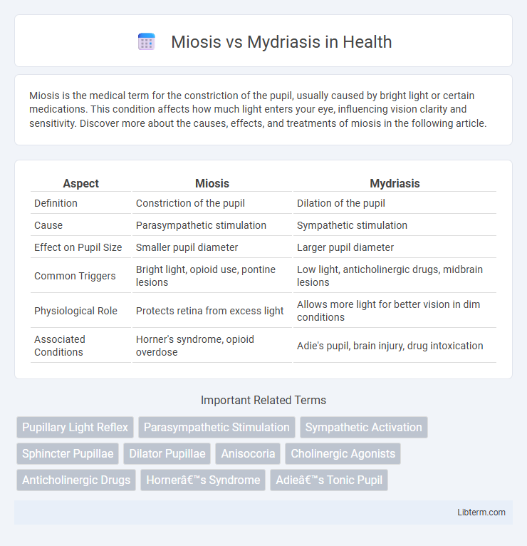
| Aspect | Miosis | Mydriasis |
|---|---|---|
| Definition | Constriction of the pupil | Dilation of the pupil |
| Cause | Parasympathetic stimulation | Sympathetic stimulation |
| Effect on Pupil Size | Smaller pupil diameter | Larger pupil diameter |
| Common Triggers | Bright light, opioid use, pontine lesions | Low light, anticholinergic drugs, midbrain lesions |
| Physiological Role | Protects retina from excess light | Allows more light for better vision in dim conditions |
| Associated Conditions | Horner's syndrome, opioid overdose | Adie's pupil, brain injury, drug intoxication |
Introduction to Miosis and Mydriasis
Miosis refers to the constriction of the pupil, reducing its diameter due to the activation of the sphincter pupillae muscle, which is controlled by the parasympathetic nervous system. Mydriasis is the dilation of the pupil, increasing its size through the contraction of the dilator pupillae muscle, influenced primarily by the sympathetic nervous system. Both pupillary responses play crucial roles in regulating the amount of light entering the eye and are essential for visual adaptation under varying lighting conditions.
Definitions: What Are Miosis and Mydriasis?
Miosis is the medical term for constriction of the pupil, typically resulting from parasympathetic nervous system activation or exposure to bright light. Mydriasis refers to the dilation of the pupil, often caused by sympathetic nervous system stimulation, low light conditions, or certain medications like anticholinergics. Both conditions affect the amount of light entering the eye, playing crucial roles in vision adaptation and diagnostic evaluations.
Anatomy of the Pupil: How Size is Controlled
The pupil size is controlled by the iris, consisting of two main muscles: the sphincter pupillae and the dilator pupillae. Miosis occurs when the sphincter pupillae contracts, narrowing the pupil and reducing light entry, primarily under parasympathetic nervous system control. Mydriasis results from the dilator pupillae muscle contracting, enlarging the pupil to increase light intake, regulated by the sympathetic nervous system.
Causes of Miosis
Miosis, characterized by the excessive constriction of the pupil, results primarily from increased parasympathetic stimulation affecting the sphincter pupillae muscle. Common causes include exposure to opioid drugs, pontine hemorrhage, and eye conditions such as iritis or glaucoma. Unlike mydriasis, which involves pupil dilation due to sympathetic activation or anticholinergic agents, miosis often indicates underlying neurological or pharmacological influences affecting parasympathetic pathways.
Causes of Mydriasis
Mydriasis, characterized by the dilation of the pupil, is primarily caused by sympathetic nervous system activation or parasympathetic inhibition affecting the iris dilator muscle. Common causes include the use of anticholinergic drugs such as atropine, central nervous system injuries, and certain recreational substances like cocaine or amphetamines. Neurological disorders including Adie's pupil and third cranial nerve palsy also contribute to pathological mydriasis.
Symptoms and Clinical Presentation
Miosis presents with abnormally constricted pupils, often less than 2 mm in diameter, accompanied by reduced vision in low light and potential eye discomfort. Mydriasis is characterized by excessive pupil dilation, typically greater than 6 mm, leading to light sensitivity, blurred vision, and difficulty focusing on near objects. Both conditions can indicate underlying neurological or ocular disorders and require thorough clinical evaluation for accurate diagnosis.
Diagnostic Approaches
Diagnostic approaches for miosis involve assessing pupillary size reduction typically through slit-lamp examination and pharmacological testing with agents like pilocarpine to determine parasympathetic overactivity or nerve damage. Mydriasis diagnosis includes evaluating pupil dilation using pupillometry, neuroimaging to rule out cranial nerve III palsy or brain injury, and administration of sympathomimetic or anticholinergic drugs to distinguish underlying causes. Both conditions require careful neurological examination to identify systemic or localized disorders affecting the autonomic nervous system.
Miosis vs Mydriasis: Key Differences
Miosis and mydriasis are opposite pupillary responses controlled by the autonomic nervous system, with miosis causing pupil constriction due to parasympathetic activation and mydriasis causing pupil dilation through sympathetic stimulation. Miosis can indicate conditions such as opioid use or brainstem injury, while mydriasis often signals sympathetic overactivity, trauma, or drug effects like anticholinergics. Understanding these differences is crucial in clinical diagnosis, aiding in the assessment of neurological function and potential underlying pathologies.
Treatment Options and Management
Treatment options for miosis focus on addressing underlying causes such as opioid overdose with naloxone administration to reverse excessive pupil constriction. Mydriasis treatment involves managing symptoms and causes like anticholinergic toxicity, often using physostigmine or other antidotes to reduce pupil dilation. Both conditions require careful monitoring of neurological status and adjustment of medications to restore normal autonomic control of the pupil.
Preventive Strategies and Patient Education
Preventive strategies for miosis and mydriasis include protecting the eyes from excessive light exposure using sunglasses with UV protection and avoiding prolonged use of medications that can trigger abnormal pupil constriction or dilation, such as opioids or anticholinergics. Patient education emphasizes the importance of recognizing early symptoms, maintaining scheduled ophthalmologic evaluations, and understanding the impact of systemic diseases like diabetes or neurological disorders on pupil function. Encouraging patients to report sudden changes in pupil size or vision disturbances helps in timely diagnosis and management, reducing the risk of complications like glaucoma or optic nerve damage.
Miosis Infographic