A ranula is a mucous cyst that forms in the floor of the mouth, often caused by the obstruction of salivary glands. This condition can lead to swelling and discomfort, sometimes interfering with speaking or eating. Discover more about symptoms, causes, and effective treatments by reading the full article.
Table of Comparison
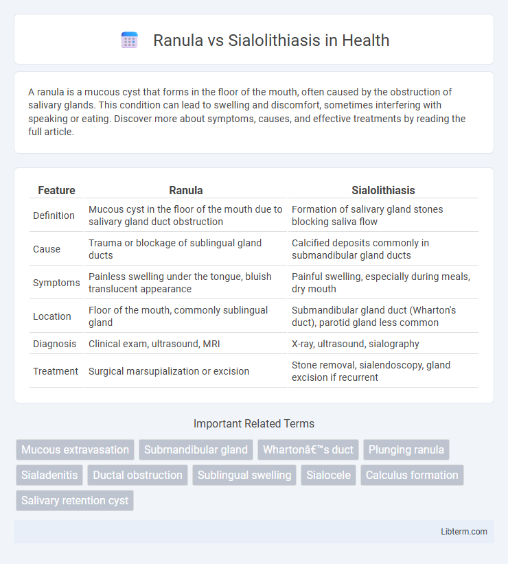
| Feature | Ranula | Sialolithiasis |
|---|---|---|
| Definition | Mucous cyst in the floor of the mouth due to salivary gland duct obstruction | Formation of salivary gland stones blocking saliva flow |
| Cause | Trauma or blockage of sublingual gland ducts | Calcified deposits commonly in submandibular gland ducts |
| Symptoms | Painless swelling under the tongue, bluish translucent appearance | Painful swelling, especially during meals, dry mouth |
| Location | Floor of the mouth, commonly sublingual gland | Submandibular gland duct (Wharton's duct), parotid gland less common |
| Diagnosis | Clinical exam, ultrasound, MRI | X-ray, ultrasound, sialography |
| Treatment | Surgical marsupialization or excision | Stone removal, sialendoscopy, gland excision if recurrent |
Introduction to Salivary Gland Disorders
Salivary gland disorders primarily include ranula and sialolithiasis, both affecting saliva flow but differing in etiology and presentation. Ranula is a mucous cyst arising from the sublingual gland due to ductal obstruction or rupture, characterized by swelling in the floor of the mouth. Sialolithiasis involves the formation of calcified stones within salivary ducts, most commonly in the submandibular gland, leading to pain and swelling during salivation.
What is a Ranula?
A ranula is a mucous cyst that forms on the floor of the mouth due to the obstruction or rupture of the sublingual gland's salivary ducts, leading to saliva accumulation in the surrounding tissues. It presents as a painless, translucent, bluish swelling that can vary in size and may cause discomfort or difficulty in speaking and swallowing if large. Unlike sialolithiasis, which involves the formation of calcified stones within salivary glands causing pain and swelling during meals, a ranula results from mucus extravasation without stone formation.
What is Sialolithiasis?
Sialolithiasis is the formation of calcified stones within the salivary glands or ducts, most commonly affecting the submandibular gland due to its mucous-rich saliva and duct anatomy. These salivary stones cause ductal obstruction, leading to pain, swelling, and sometimes infection during meals. Unlike ranulas, which are mucous cysts caused by ductal rupture or obstruction, sialolithiasis specifically involves the physical blockage by mineralized calculi disrupting saliva flow.
Etiology: Causes of Ranula vs Sialolithiasis
Ranula primarily arises from the obstruction or trauma to the sublingual salivary gland ducts, leading to mucus extravasation and cyst formation. Sialolithiasis is caused by the formation of calcified concretions or salivary stones within the salivary gland ducts, frequently the submandibular gland, due to factors like saliva stasis, altered saliva composition, or ductal injury. The differing etiologies distinguish ranulas as mucus retention cysts and sialolithiasis as obstructive conditions due to mineralized deposits.
Clinical Presentation: Signs and Symptoms
Ranula typically presents as a painless, bluish, translucent swelling on the floor of the mouth, often causing a visible bulge under the tongue and occasional difficulty swallowing or speaking. Sialolithiasis manifests with recurrent pain and swelling in the affected salivary gland, especially during meals, accompanied by possible infection or pus discharge if obstruction persists. Both conditions can cause gland enlargement, but ranula is primarily cystic, whereas sialolithiasis involves obstructive sialadenitis related to salivary duct stones.
Diagnostic Approaches and Imaging
Diagnostic approaches for Ranula primarily involve clinical examination and ultrasound imaging to identify cystic lesions originating from the salivary glands, while sialography and CT scans are more effective for detecting Sialolithiasis caused by mineralized salivary stones. MRI provides detailed soft tissue contrast useful in distinguishing ranulas from other cystic masses and can assess the extent of fluid collection, whereas non-contrast CT excels in identifying calcified sialoliths within the salivary ducts. Ultrasonography, due to its accessibility and lack of radiation, serves as a first-line imaging modality in both conditions but requires correlation with clinical findings for accurate differential diagnosis.
Key Differences in Pathophysiology
Ranula is a mucous extravasation cyst originating from the sublingual gland, caused by trauma or obstruction of the salivary gland duct resulting in mucus accumulation in the floor of the mouth. Sialolithiasis involves the formation of calcified sialoliths or salivary stones, primarily in the submandibular gland duct, leading to ductal obstruction and subsequent glandular swelling and pain. Pathophysiologically, ranula results from mucus leakage and cyst formation due to ductal rupture, whereas sialolithiasis is characterized by mineral deposition causing mechanical blockage and inflammatory changes.
Treatment Modalities: Ranula vs Sialolithiasis
Treatment modalities for ranula primarily involve marsupialization or complete excision of the cyst along with the associated sublingual gland to prevent recurrence. In contrast, sialolithiasis treatment focuses on removing the salivary gland stones through sialolithotomy, sialendoscopy, or gland excision in severe cases. Minimally invasive sialendoscopy offers an effective approach for sialolithiasis with less morbidity compared to traditional surgery.
Complications and Prognosis
Ranulas, arising from mucous extravasation in the floor of the mouth, often lead to airway obstruction or infection if untreated, whereas sialolithiasis, characterized by salivary gland duct stones, primarily causes recurrent gland swelling and secondary bacterial infections. Complications of ranulas include potential cyst rupture and abscess formation, while sialolithiasis may result in chronic sialadenitis or gland atrophy. The prognosis for ranulas improves with surgical marsupialization but carries a risk of recurrence; sialolithiasis prognosis depends on stone removal success, with early intervention preventing chronic gland damage.
Prevention and Patient Education
Ranulas primarily result from trauma or obstruction of the sublingual gland duct, making avoidance of oral injuries and maintaining good oral hygiene crucial for prevention. Sialolithiasis, caused by calcified stones in salivary glands, can be minimized by staying well-hydrated, massaging glands regularly, and avoiding medications that reduce saliva flow. Patient education should emphasize recognizing symptoms early, such as swelling and pain, and seeking prompt dental or medical evaluation to prevent complications.
Ranula Infographic