Sialadenitis is an inflammation of the salivary glands often caused by bacterial or viral infections, leading to pain, swelling, and sometimes fever. Prompt diagnosis and treatment are crucial to prevent complications such as abscess formation or gland dysfunction. Explore the rest of the article to understand symptoms, causes, and effective treatment options for your condition.
Table of Comparison
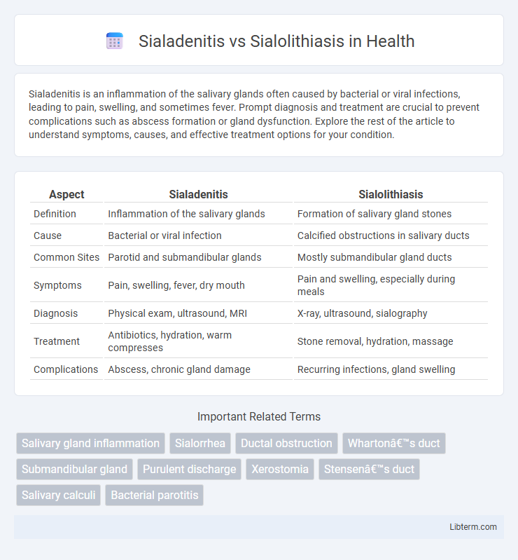
| Aspect | Sialadenitis | Sialolithiasis |
|---|---|---|
| Definition | Inflammation of the salivary glands | Formation of salivary gland stones |
| Cause | Bacterial or viral infection | Calcified obstructions in salivary ducts |
| Common Sites | Parotid and submandibular glands | Mostly submandibular gland ducts |
| Symptoms | Pain, swelling, fever, dry mouth | Pain and swelling, especially during meals |
| Diagnosis | Physical exam, ultrasound, MRI | X-ray, ultrasound, sialography |
| Treatment | Antibiotics, hydration, warm compresses | Stone removal, hydration, massage |
| Complications | Abscess, chronic gland damage | Recurring infections, gland swelling |
Introduction to Salivary Gland Disorders
Salivary gland disorders primarily include sialadenitis, characterized by inflammation, and sialolithiasis, marked by the formation of salivary stones. Sialadenitis results from infections or autoimmune conditions leading to gland swelling and pain, while sialolithiasis involves mineralized obstructions that cause ductal blockage and saliva flow impairment. Understanding the differences between these conditions is critical for accurate diagnosis and effective treatment of salivary gland dysfunctions.
Defining Sialadenitis
Sialadenitis is an inflammation of the salivary glands caused by bacterial or viral infections, often leading to swelling, pain, and sometimes pus drainage. It primarily affects the parotid or submandibular glands and can result from ductal obstruction, dehydration, or immunosuppression. Sialolithiasis, by contrast, involves the formation of salivary stones that block gland ducts, causing pain and swelling but is distinguished from Sialadenitis by its mechanical obstruction rather than infectious inflammation.
What is Sialolithiasis?
Sialolithiasis is the formation of calcified stones within the salivary glands or their ducts, most commonly affecting the submandibular gland due to its viscous saliva and upward duct flow. These stones can cause obstruction, leading to swelling, pain, and sometimes infection if saliva flow is blocked. Treatment may involve hydration, massage, lithotripsy, or surgical removal depending on the size and location of the calculi.
Key Differences Between Sialadenitis and Sialolithiasis
Sialadenitis is an inflammation of the salivary glands often caused by bacterial or viral infections, whereas sialolithiasis refers to the formation of calcified stones blocking the salivary ducts. Sialadenitis typically presents with gland swelling, pain, and possible pus discharge, while sialolithiasis primarily causes intermittent pain and swelling, especially during meals due to saliva blockage. Diagnostic imaging such as ultrasound or sialography helps differentiate sialadenitis, characterized by gland inflammation, from sialolithiasis, identified by visible ductal calculi.
Causes and Risk Factors of Sialadenitis
Sialadenitis is primarily caused by bacterial infections, often due to Staphylococcus aureus, with reduced salivary flow creating an environment conducive to microbial growth. Risk factors include dehydration, Sjogren's syndrome, ductal obstruction, and immunosuppression, all contributing to saliva stasis and inflammation. In contrast, sialolithiasis arises from salivary gland duct stones causing mechanical blockage and subsequent gland swelling.
Etiology and Pathogenesis of Sialolithiasis
Sialolithiasis is primarily caused by the formation of calcified deposits within the salivary gland ducts, most commonly affecting the submandibular gland due to its alkaline saliva and slower salivary flow. The pathogenesis involves precipitation of calcium salts around an initial organic nidus, often composed of mucus, bacteria, or desquamated epithelial cells, leading to ductal obstruction and subsequent glandular swelling and pain. Unlike sialadenitis, which is an inflammation typically resulting from infection or autoimmune causes, sialolithiasis directly causes mechanical blockage that predisposes the gland to secondary infection and inflammation.
Common Symptoms and Clinical Presentation
Sialadenitis typically presents with painful swelling of the affected salivary gland, often accompanied by fever, erythema, and purulent discharge from the duct. Sialolithiasis commonly manifests as episodic pain and swelling in the salivary glands, especially during meals, due to obstruction from salivary stones. Both conditions cause gland enlargement and tenderness, but sialadenitis is more associated with infection signs, whereas sialolithiasis primarily involves mechanical blockage symptoms.
Diagnostic Strategies and Imaging Techniques
Sialadenitis is primarily diagnosed through clinical examination and confirmed with ultrasound imaging, which reveals gland enlargement and hypoechoic areas indicative of inflammation. Sialolithiasis diagnosis relies heavily on imaging modalities such as non-contrast computed tomography (CT) and sialography to detect calcified stones within salivary ducts. Magnetic resonance sialography (MR sialography) offers a non-invasive method to evaluate ductal system obstructions and differentiate between inflammatory changes in sialadenitis and obstructive stones in sialolithiasis.
Treatment Options and Management Approaches
Sialadenitis treatment primarily involves antibiotics targeting bacterial infections, along with hydration and gland massage to promote saliva flow, while severe cases may require surgical drainage. Sialolithiasis management focuses on stone removal, either through manual expression, sialogogues to stimulate saliva, or minimally invasive procedures such as sialendoscopy; larger or obstructive stones can necessitate surgical excision. Both conditions benefit from pain control measures and preventive strategies to maintain salivary gland function and reduce recurrence risk.
Prevention, Prognosis, and Patient Education
Sialadenitis prevention involves maintaining optimal oral hygiene and adequate hydration to reduce bacterial infection risks, whereas sialolithiasis prevention emphasizes regular massage and stimulation of salivary flow to avoid stone formation. Prognosis for sialadenitis improves with timely antibiotic therapy, while sialolithiasis prognosis depends on stone size and location, where small stones often resolve with conservative measures, but larger stones may require surgical removal. Patient education should focus on recognizing early symptoms such as gland swelling and pain, advising prompt medical evaluation, and encouraging avoidance of dehydration and behaviors that reduce salivary flow.
Sialadenitis Infographic