Basophilic cells stain readily with basic dyes due to their affinity for acidic components such as nucleic acids, playing a crucial role in histological analysis and diagnosis. Understanding basophilic properties can help you interpret tissue samples more accurately and identify pathological changes. Explore the article to learn more about the significance and applications of basophilic staining in medical science.
Table of Comparison
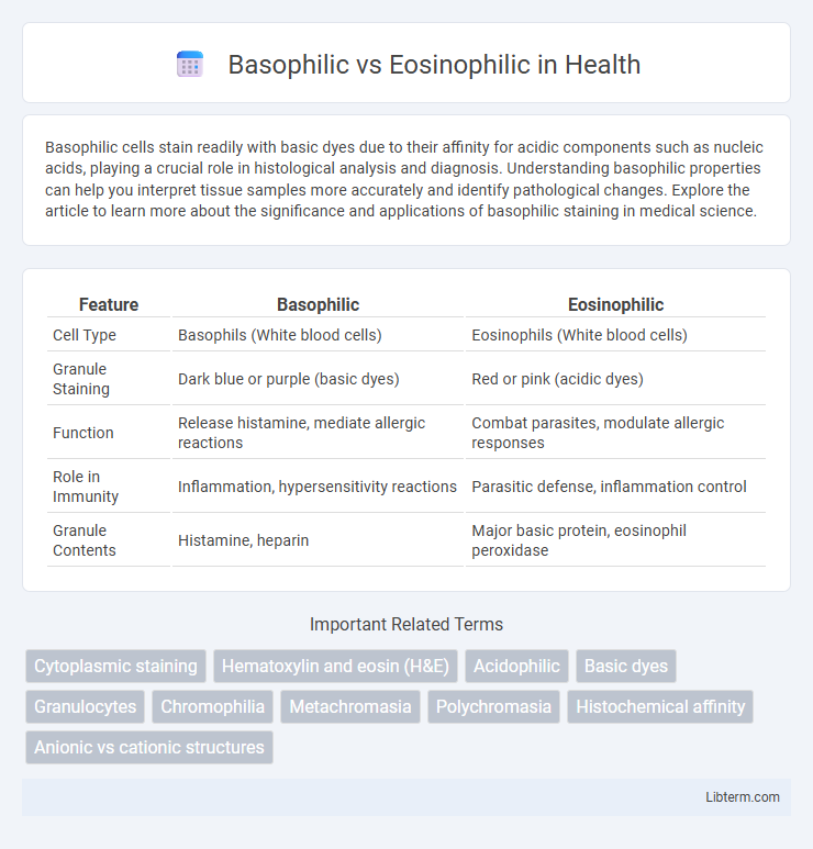
| Feature | Basophilic | Eosinophilic |
|---|---|---|
| Cell Type | Basophils (White blood cells) | Eosinophils (White blood cells) |
| Granule Staining | Dark blue or purple (basic dyes) | Red or pink (acidic dyes) |
| Function | Release histamine, mediate allergic reactions | Combat parasites, modulate allergic responses |
| Role in Immunity | Inflammation, hypersensitivity reactions | Parasitic defense, inflammation control |
| Granule Contents | Histamine, heparin | Major basic protein, eosinophil peroxidase |
Introduction to Basophilic and Eosinophilic Cells
Basophilic cells are characterized by their affinity for basic dyes, staining dark blue or purple due to the presence of acidic components like nucleic acids in the cytoplasm. Eosinophilic cells exhibit a strong affinity for acidic dyes, resulting in a pink or red appearance, mainly because of abundant proteins and granules in the cytoplasm. These staining properties are essential for identifying cell types and understanding their roles in immune response and tissue function.
Defining Basophilic: Characteristics and Staining
Basophilic cells exhibit an affinity for basic dyes such as hematoxylin, staining dark blue or purple due to the presence of acidic components like nucleic acids in the cytoplasm and nucleus. These cells play critical roles in inflammatory and allergic responses, often identified by their coarse granules rich in histamine and heparin. The basophilic staining pattern is essential in histology for distinguishing cell types in tissues, particularly in bone marrow and blood smears.
Understanding Eosinophilic: Features and Staining Patterns
Eosinophilic cells and tissues exhibit strong affinity for eosin, a red or pink acidic dye that binds to basic components like cytoplasmic proteins, highlighting structures such as mitochondria and collagen fibers with vivid pink staining. In contrast, basophilic cells and tissues stain deeply blue or purple with basic dyes like hematoxylin, indicating the presence of acidic components like nucleic acids in the nucleus and rough endoplasmic reticulum. Understanding eosinophilic staining patterns aids in identifying cytoplasmic elements and extracellular matrix proteins, crucial for histopathological analysis and diagnosing conditions involving tissue damage or inflammation.
Morphological Differences: Basophils vs. Eosinophils
Basophils exhibit large, dark purple granules that often obscure the bilobed nucleus, while eosinophils contain bright red-orange, uniform granules with a distinct bilobed nucleus visible. Basophilic granules are rich in histamine and heparin, contributing to allergic reactions and inflammation, whereas eosinophilic granules contain major basic protein and eosinophil peroxidase, which are critical for combating parasitic infections. Morphologically, basophils are typically less abundant, approximately 0.5-1% of circulating leukocytes, compared to eosinophils, which constitute about 1-4% and have slightly larger cell size.
Functional Roles in the Immune System
Basophilic cells, primarily basophils and mast cells, release histamine and other mediators to initiate inflammation and allergic responses, enhancing vascular permeability and recruiting other immune cells. Eosinophilic cells, mainly eosinophils, play a critical role in combating parasitic infections and modulating allergic inflammation by releasing cytotoxic granules and cytokines. Both cell types contribute to immune regulation but target distinct pathogens and inflammatory pathways to maintain immune homeostasis.
Associated Diseases: Basophilia vs. Eosinophilia
Basophilia is commonly associated with chronic myelogenous leukemia (CML), allergic reactions, and autoimmune disorders, where elevated basophil counts contribute to inflammation and histamine release. Eosinophilia is frequently linked to parasitic infections, allergic conditions such as asthma and eczema, and certain malignancies like Hodgkin's lymphoma, characterized by increased eosinophil activity in tissue damage and immune response. Both conditions involve distinct pathophysiological mechanisms reflected in their differential roles in immune regulation and disease processes.
Diagnostic Importance in Laboratory Analysis
Basophilic and eosinophilic staining are critical in laboratory diagnostics for differentiating cell types and identifying pathological changes. Basophilic cells, which stain blue or purple due to affinity for basic dyes, are often associated with nucleic acids and indicate lymphoid tissues or inflammation. Eosinophilic cells, staining pink or red with acidic dyes, highlight cytoplasmic components and are essential for diagnosing allergic reactions, parasitic infections, and certain hematologic disorders.
Histological Appearance in Tissue Samples
Basophilic cells exhibit a strong affinity for basic dyes, appearing deep blue or purple in histological tissue samples due to their abundant nucleic acids and acidic components. Eosinophilic cells stain vividly with eosin dye, showing a bright pink or red color caused by their high protein content and abundant cytoplasmic granules. The contrast in staining patterns between basophilic and eosinophilic cells aids in distinguishing cell types and identifying tissue pathology under microscopy.
Clinical Implications and Treatment Considerations
Basophilic and eosinophilic cells differ in their staining properties and immune functions, with basophils primarily involved in allergic responses and eosinophils playing key roles in parasitic infections and asthma. Clinically, elevated basophil counts are often associated with chronic myelogenous leukemia and hypersensitivity disorders, while increased eosinophils indicate allergic conditions, parasitic infections, or eosinophilic esophagitis. Treatment considerations vary accordingly, with basophilic-driven conditions managed using antihistamines or corticosteroids to control histamine release, whereas eosinophilic disorders may require targeted therapies like anti-IL-5 monoclonal antibodies to reduce eosinophil activity and inflammation.
Summary: Basophilic vs. Eosinophilic Comparison
Basophilic cells stain deeply with basic dyes due to their affinity for acidic components like DNA and RNA, highlighting structures such as nuclei and ribosomes, whereas eosinophilic cells stain strongly with acidic dyes like eosin, indicating a high concentration of basic proteins, typically found in cytoplasmic granules. Basophilia is characteristic of basophils, mast cells, and certain cell types rich in nucleic acids, while eosinophilia is prominent in eosinophils and cells with abundant mitochondria or secretory granules. Understanding these staining differences is crucial for histological identification and diagnosing various pathological conditions.
Basophilic Infographic