Spherocytosis is a hereditary condition characterized by abnormally shaped red blood cells that appear spherical instead of the typical biconcave disc shape, leading to their premature destruction and resulting in anemia. Symptoms often include fatigue, jaundice, and an enlarged spleen, which can affect Your overall health and energy levels. Explore the rest of this article to understand the causes, symptoms, and treatment options for managing spherocytosis effectively.
Table of Comparison
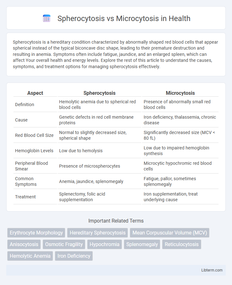
| Aspect | Spherocytosis | Microcytosis |
|---|---|---|
| Definition | Hemolytic anemia due to spherical red blood cells | Presence of abnormally small red blood cells |
| Cause | Genetic defects in red cell membrane proteins | Iron deficiency, thalassemia, chronic disease |
| Red Blood Cell Size | Normal to slightly decreased size, spherical shape | Significantly decreased size (MCV < 80 fL) |
| Hemoglobin Levels | Low due to hemolysis | Low due to impaired hemoglobin synthesis |
| Peripheral Blood Smear | Presence of microspherocytes | Microcytic hypochromic red blood cells |
| Common Symptoms | Anemia, jaundice, splenomegaly | Fatigue, pallor, sometimes splenomegaly |
| Treatment | Splenectomy, folic acid supplementation | Iron supplementation, treat underlying cause |
Overview of Spherocytosis and Microcytosis
Spherocytosis is a hereditary condition characterized by the presence of sphere-shaped red blood cells, leading to hemolytic anemia due to their reduced deformability and premature destruction in the spleen. Microcytosis refers to the presence of abnormally small red blood cells, often associated with iron deficiency anemia or thalassemia, which impairs oxygen transport capacity. Both disorders affect red blood cell morphology but differ fundamentally in their pathophysiology and clinical implications.
Definition and Key Characteristics
Spherocytosis is a hereditary blood disorder characterized by the presence of spherical red blood cells that lack the usual biconcave shape, leading to increased cell fragility and hemolytic anemia. Microcytosis refers to the presence of abnormally small red blood cells, often associated with conditions such as iron deficiency anemia or thalassemia. Key characteristics of spherocytosis include hyperchromic, normocytic red cells with reduced membrane surface area, while microcytosis features hypochromic, microcytic red cells with decreased hemoglobin content.
Causes and Underlying Mechanisms
Spherocytosis results from genetic mutations affecting ankyrin, spectrin, or band 3 proteins, causing red blood cells to lose their typical biconcave shape and become spherical, leading to premature splenic destruction. Microcytosis is primarily caused by disorders like iron deficiency anemia or thalassemia, where impaired hemoglobin synthesis results in smaller-than-normal red blood cells. The underlying mechanisms in spherocytosis involve membrane protein defects disrupting cytoskeletal stability, while microcytosis stems from defective hemoglobin production reducing cell size.
Clinical Manifestations
Spherocytosis typically presents with anemia, jaundice, splenomegaly, and increased reticulocyte count due to hemolysis caused by spherical red blood cells. Microcytosis, often associated with iron deficiency anemia or thalassemia, manifests as fatigue, pallor, and microcytic hypochromic red blood cells on peripheral smear. Both conditions exhibit distinct hematologic profiles essential for differential diagnosis in clinical practice.
Diagnostic Criteria and Laboratory Findings
Spherocytosis is characterized by the presence of spherical red blood cells on peripheral blood smear, increased mean corpuscular hemoglobin concentration (MCHC), and a positive osmotic fragility test or eosin-5'-maleimide (EMA) binding test. Microcytosis presents with decreased mean corpuscular volume (MCV) and is commonly associated with iron deficiency anemia or thalassemia, confirmed by low serum ferritin or hemoglobin electrophoresis, respectively. Both conditions show distinct laboratory patterns: spherocytosis with normocytic or slightly microcytic indices and increased reticulocyte count, while microcytosis demonstrates uniformly small red cells and variable anisopoikilocytosis.
Morphological Differences in Red Blood Cells
Spherocytosis is characterized by the presence of spherical, dense red blood cells lacking the normal central pallor, resulting in decreased membrane surface area and increased osmotic fragility. Microcytosis features abnormally small red blood cells with a reduced mean corpuscular volume (MCV), often accompanied by hypochromia and pronounced central pallor due to decreased hemoglobin content. Morphological evaluation differentiates spherocytes' round, dense appearance from microcytes' small size and pale coloration on peripheral blood smear.
Genetic and Acquired Forms
Spherocytosis primarily results from genetic mutations affecting proteins like ankyrin, spectrin, and band 3, leading to hereditary spherocytosis characterized by rigid, spherical red blood cells. Microcytosis can arise from genetic causes such as thalassemia mutations affecting hemoglobin synthesis or acquired conditions like iron deficiency anemia, influencing red blood cell size reduction. Understanding the distinct genetic mutations and acquired factors underlying spherocytosis and microcytosis is critical for accurate diagnosis and tailored treatment strategies.
Treatment and Management Strategies
Spherocytosis treatment primarily involves folic acid supplementation, blood transfusions during severe anemia, and splenectomy to reduce hemolysis in hereditary cases. Microcytosis management depends on the underlying cause, often requiring iron supplementation for iron-deficiency anemia or specific therapies for thalassemia and other hemoglobinopathies. Regular monitoring of blood counts and tailored interventions based on genetic testing and iron studies optimize patient outcomes in both conditions.
Prognosis and Long-Term Outcomes
Hereditary spherocytosis typically has a favorable prognosis with proper management, including folic acid supplementation and, in severe cases, splenectomy, which significantly improves quality of life and reduces hemolytic episodes. In contrast, microcytosis, often caused by iron deficiency anemia or thalassemia, requires ongoing treatment tailored to the underlying cause, with iron supplementation or genetic counseling impacting long-term outcomes. Both conditions necessitate regular monitoring for complications like anemia severity and organ damage to ensure optimal patient prognosis.
Summary Table: Spherocytosis vs Microcytosis
Spherocytosis is characterized by the presence of spherical red blood cells with reduced membrane surface area, leading to hemolytic anemia, whereas microcytosis involves abnormally small red blood cells often associated with iron deficiency or thalassemia. Key diagnostic features include increased osmotic fragility and positive family history in spherocytosis, contrasted with low mean corpuscular volume (MCV) and hypochromia in microcytosis. Laboratory tests such as peripheral blood smear, osmotic fragility test for spherocytosis, and iron studies or hemoglobin electrophoresis for microcytosis are essential for accurate differentiation.
Spherocytosis Infographic