Empyema, a collection of pus in the pleural cavity, can complicate lung cancer by causing infections and impairing lung function. Managing empyema alongside lung cancer requires careful diagnosis and treatment to prevent further respiratory compromise and improve patient outcomes. Explore the full article to understand how these conditions interact and what treatment options might be best for you.
Table of Comparison
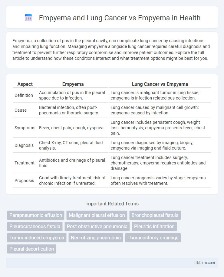
| Aspect | Empyema | Lung Cancer vs Empyema |
|---|---|---|
| Definition | Accumulation of pus in the pleural space due to infection. | Lung cancer is malignant tumor in lung tissue; empyema is infection-related pus collection. |
| Cause | Bacterial infection, often post-pneumonia or thoracic surgery. | Lung cancer caused by malignant cell growth; empyema caused by infection. |
| Symptoms | Fever, chest pain, cough, dyspnea. | Lung cancer includes persistent cough, weight loss, hemoptysis; empyema presents fever, chest pain. |
| Diagnosis | Chest X-ray, CT scan, pleural fluid analysis. | Lung cancer diagnosed by imaging, biopsy; empyema via imaging and fluid culture. |
| Treatment | Antibiotics and drainage of pleural fluid. | Lung cancer treatment includes surgery, chemotherapy; empyema requires antibiotics and drainage. |
| Prognosis | Good with timely treatment; risk of chronic infection if untreated. | Lung cancer prognosis varies by stage; empyema often resolves with treatment. |
Introduction to Empyema
Empyema is a collection of pus in the pleural cavity, often resulting from bacterial pneumonia, lung abscess, or thoracic surgery complications. This condition causes inflammation and infection, leading to thickened pleural membranes and impaired lung function. In contrast, empyema associated with lung cancer may involve malignant pleural effusions complicated by secondary infection, complicating diagnosis and treatment.
Overview of Lung Cancer
Lung cancer is a malignant tumor characterized by uncontrolled cell growth in lung tissues, often linked to smoking and environmental factors, leading to symptoms such as persistent cough, hemoptysis, and weight loss. Empyema, an accumulation of pus in the pleural space usually caused by infection, can sometimes complicate lung cancer due to tumor-induced obstruction or secondary infections. Differentiating between empyema and lung cancer involves imaging techniques like CT scans and biopsies to accurately diagnose and guide appropriate treatment strategies.
Pathophysiology of Empyema
Empyema is characterized by the accumulation of pus within the pleural cavity, primarily resulting from bacterial infections that lead to inflammation, fibrin deposition, and pleural thickening. In cases complicated by lung cancer, tumor invasion disrupts normal pleural integrity, promoting secondary infections and impaired drainage, exacerbating empyema formation. The underlying pathophysiology involves an inflammatory cascade that progresses from simple exudative effusions to fibropurulent stages, culminating in organizing phases with dense pleural adhesions and lung entrapment.
Causes of Empyema in Lung Cancer Patients
Empyema in lung cancer patients primarily arises from tumor invasion leading to bronchial obstruction, causing post-obstructive pneumonia and subsequent pleural infection. Malignant pleural effusion can become secondarily infected, contributing to empyema development. Immunosuppression due to cancer or chemotherapy increases susceptibility to pleural infections, differentiating empyema causes in lung cancer from those in otherwise healthy individuals.
Clinical Presentation: Empyema vs Empyema in Lung Cancer
Empyema typically presents with fever, pleuritic chest pain, productive cough, and dyspnea, while empyema associated with lung cancer often shows similar respiratory symptoms but may also involve systemic signs such as unintended weight loss and hemoptysis due to the underlying malignancy. Diagnostic imaging in both cases reveals pleural fluid collections, but lung cancer-related empyema frequently exhibits irregular pleural thickening or nodules suggestive of tumor invasion. Laboratory analysis of pleural fluid in empyema shows purulent effusion with high white cell count and low pH, whereas in lung cancer-associated empyema, malignant cells may be detected, indicating neoplastic involvement.
Diagnostic Strategies for Empyema and Lung Cancer-Associated Empyema
Empyema diagnosis relies on thoracic ultrasound and pleural fluid analysis to detect purulent effusion and distinguish bacterial infections from malignancies. In lung cancer-associated empyema, advanced imaging such as contrast-enhanced CT scans and PET-CT are critical for identifying tumor invasion and differentiating malignant pleural involvement. Combining microbiological cultures with cytology and biomarker analysis improves diagnostic accuracy for both infectious empyema and cancer-related pleural complications.
Imaging and Laboratory Findings
Empyema often presents on imaging with pleural fluid loculations, thickened pleural membranes, and air-fluid levels, while lung cancer typically shows a solitary pulmonary mass, possible lymphadenopathy, and possible pleural effusion on CT or chest X-ray. Laboratory analysis of pleural fluid in empyema reveals purulent fluid with low pH (<7.2), low glucose, elevated lactate dehydrogenase (LDH), and positive bacterial cultures, indicative of infection. In contrast, malignant pleural effusions associated with lung cancer exhibit serous or hemorrhagic fluid with high tumor marker levels, cytology positive for malignant cells, and less pronounced inflammatory markers compared to empyema.
Treatment Approaches: Isolated Empyema vs Empyema in Lung Cancer
Treatment of isolated empyema typically involves prompt antibiotic therapy combined with thoracic drainage to evacuate purulent fluid and prevent lung entrapment. In empyema associated with lung cancer, therapeutic strategies become more complex, often requiring surgical intervention such as decortication or tumor resection alongside infection control to address both malignant and infectious processes. The presence of malignancy necessitates multidisciplinary management to optimize outcomes, balancing oncologic treatment with aggressive infection resolution.
Prognosis and Outcomes
Empyema associated with lung cancer typically leads to a poorer prognosis due to the underlying malignancy compromising immune response and lung function, resulting in higher morbidity and mortality rates compared to empyema alone. Outcomes in empyema without malignancy often improve significantly with prompt antibiotic therapy and surgical drainage, whereas lung cancer-related empyema frequently necessitates multidisciplinary oncologic and thoracic interventions. Survival rates are considerably lower in patients with lung cancer-associated empyema, emphasizing the critical importance of early detection and integrated treatment strategies.
Preventive Measures and Future Perspectives
Preventive measures for empyema and lung cancer primarily involve reducing risk factors such as smoking cessation, early vaccination against respiratory infections, and prompt treatment of pneumonia to prevent pleural infection. Advances in diagnostic imaging and molecular profiling enhance early detection and personalized treatment plans, improving patient outcomes. Future perspectives include the development of targeted therapies for lung cancer and novel antimicrobial strategies to manage empyema, alongside improved public health initiatives to reduce incidence rates globally.
Empyema and Lung Cancer Infographic