Muscle hypertrophy is the process of increasing muscle size through targeted resistance training and proper nutrition. Maximizing hypertrophy involves understanding the balance of volume, intensity, and recovery in your workout routine. Discover effective strategies to optimize your muscle growth by exploring the rest of the article.
Table of Comparison
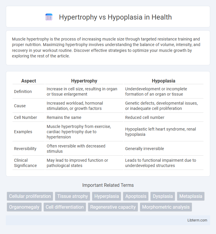
| Aspect | Hypertrophy | Hypoplasia |
|---|---|---|
| Definition | Increase in cell size, resulting in organ or tissue enlargement | Underdevelopment or incomplete formation of an organ or tissue |
| Cause | Increased workload, hormonal stimulation, or growth factors | Genetic defects, developmental issues, or inadequate cell proliferation |
| Cell Number | Remains the same | Reduced cell number |
| Examples | Muscle hypertrophy from exercise, cardiac hypertrophy due to hypertension | Hypoplastic left heart syndrome, renal hypoplasia |
| Reversibility | Often reversible with decreased stimulus | Generally irreversible |
| Clinical Significance | May lead to improved function or pathological states | Leads to functional impairment due to underdeveloped structures |
Understanding Hypertrophy and Hypoplasia
Hypertrophy refers to the increase in the size of cells, leading to the enlargement of an organ or tissue without an increase in cell number, commonly seen in muscle growth due to exercise. Hypoplasia, in contrast, describes the underdevelopment or incomplete formation of a tissue or organ resulting from a reduced number of cells during embryonic development. Understanding the distinct cellular mechanisms and developmental stages involved is crucial for diagnosing and managing conditions related to abnormal tissue growth or development.
Definitions: Hypertrophy vs Hypoplasia
Hypertrophy is the increase in the size of cells, resulting in the enlargement of an organ or tissue without an increase in cell number, commonly observed in muscles due to exercise. Hypoplasia refers to the underdevelopment or incomplete formation of an organ or tissue caused by a reduced number of cells during embryonic development. Understanding the distinction between hypertrophy and hypoplasia is critical in diagnosing developmental abnormalities and assessing tissue growth responses.
Causes of Hypertrophy
Hypertrophy occurs due to increased functional demand or hormonal stimulation, causing cells to enlarge by synthesizing more structural components. Common causes include resistance training, which induces muscle hypertrophy through mechanical stress, and hormonal factors like insulin-like growth factor 1 (IGF-1) that promote protein synthesis in tissues. Unlike hypoplasia, which stems from incomplete development and results in fewer cells, hypertrophy involves the enlargement of existing cells without an increase in cell number.
Causes of Hypoplasia
Hypoplasia results from incomplete development or underdevelopment of a tissue or organ, often due to genetic mutations, intrauterine factors like fetal malnutrition, or exposure to teratogens during critical periods of organogenesis. This condition differs from hypertrophy, which involves the enlargement of existing cells rather than a reduced number of cells caused by developmental defects. Causes of hypoplasia include chromosomal abnormalities, maternal infections such as rubella, ischemia during fetal growth, and deficiencies in essential nutrients like folic acid.
Cellular Mechanisms Involved
Hypertrophy involves an increase in cell size due to enhanced protein synthesis and organelle expansion, primarily regulated by signaling pathways such as mTOR and IGF-1/Akt. Hypoplasia is characterized by a reduced number of cells resulting from impaired cell proliferation or differentiation during development, often linked to disruptions in growth factors like FGFs and transcription factors such as Pax genes. Understanding these cellular mechanisms is crucial for distinguishing between tissue growth due to cell enlargement versus cell number deficits.
Clinical Examples of Hypertrophy
Hypertrophy refers to the increase in the size of cells leading to the enlargement of an organ, commonly seen in physiological conditions like skeletal muscle growth from exercise or pathological states such as cardiac hypertrophy due to hypertension. Clinical examples include left ventricular hypertrophy in patients with chronic high blood pressure and benign prostatic hypertrophy causing urinary symptoms in older men. Unlike hypoplasia, where there is an underdevelopment or incomplete development of a tissue or organ, hypertrophy involves an increase in existing cell size without an increase in cell number.
Clinical Examples of Hypoplasia
Hypoplasia refers to the underdevelopment or incomplete development of a tissue or organ, often resulting from inadequate cell proliferation during fetal growth. Clinical examples of hypoplasia include renal hypoplasia, characterized by fewer nephrons leading to reduced kidney function, and enamel hypoplasia, where tooth enamel is poorly formed, increasing susceptibility to dental caries. Other notable instances are pulmonary hypoplasia seen in congenital diaphragmatic hernia, resulting in insufficient lung development, and optic nerve hypoplasia causing visual impairment due to reduced nerve fibers.
Diagnostic Differences
Hypertrophy involves the enlargement of existing cells, resulting in increased organ or tissue size without new cell formation, whereas hypoplasia refers to the underdevelopment or incomplete formation of an organ or tissue due to a reduced number of cells. Diagnostic differences include imaging techniques such as ultrasound or MRI, which reveal increased tissue mass in hypertrophy and smaller-than-normal organ size in hypoplasia. Histopathological analysis further differentiates by showing enlarged cell size in hypertrophy and decreased cellularity or immature tissue architecture in hypoplasia.
Treatment and Management Approaches
Treatment and management of hypertrophy primarily involve addressing the underlying cause, such as reducing workload through physical therapy or pharmacological interventions to decrease muscle or organ size. Hypoplasia management often requires supportive care, including surgical correction or regenerative therapies to stimulate tissue growth and improve function. Monitoring progression with imaging and functional assessments guides tailored therapeutic strategies to optimize patient outcomes.
Prognosis and Outcomes
Hypertrophy involves the enlargement of existing cells, often leading to increased organ or tissue size without cell number changes, and generally presents a better prognosis if the underlying cause is controlled. Hypoplasia refers to the incomplete development or underdevelopment of a tissue or organ due to reduced cell numbers, often resulting in permanent functional deficits and a more guarded long-term outcome. Recovery potential in hypertrophy exceeds that in hypoplasia, where prognosis depends largely on the extent of cellular deficit and potential for compensatory growth.
Hypertrophy Infographic